Field of Science

The evolution of mutualistic relationships

ResearchBlogging.orgAlthough bacteria are often thought of as invading pathogens, not all bacterial interactions are necessarily to the detriment of the host. Some bacteria are able to establish mutualistic relationships which benefit both the bacteria and the organism in which it lives. An example can be seen in legumes, which have bacteria in specialised root nodules which carry out nitrogen fixation. The plant gains a source of nitrogen while the bacterium gains a safe space to live and a good source of carbohydrates.
Root of a legume, showing the bumpy nodules where the bacteria live

Studies of how bacteria evolve from free-living organisms into mutualistic partners of eukaryotic (i.e not bacterial) cells has been made easier with the availability of large numbers of sequenced genomes. Intracellular bacteria in general tend to have a smaller genome than free-living bacteria as they have fewer changing environmental conditions to respond too, and many of their metabolic needs can be met by their host. Examining bacteria within their natural environment (rather than the very specific and controlled laboratory environment) also helps to identify the selective forces that can act on bacteria to drive them towards adaptations for mutualistic living.

In order to adapt to a mutualistic lifestyle the bacteria must also gain new genes, which produce proteins that allow it to invade, and communicate with, its host. Genes involved in host interactions are often found in genomic islands, or near to mobile elements which are able to move the genes between organisms (particularly bacteriophages – virus’s that invade bacteria). It has been suggested that the ability of these mobile elements to leave one bacteria and invade neighbouring ones can help to prevent bacterial ‘cheaters’ i.e those that reap the benefits of the mutualistic relationship while relying on the surrounding bacteria to produce the necessary proteins.

Although several species have been found that are thought to have moved from being mutual partners to free-living, the reduction in genome size and host dependency means that once a relationship with the host is established, it tends to remain (occasionally breaking down into parasitism). In fact, once becoming so established that the host carried part (or all) of the bacterial genome, mutualist relationships can often evolve into organellar relationships, where the bacteria become part of the surrounding host cell, and is unable to survive on its own. Established mutualistic bacteria often lose the mobile elements that helped them establish in the first place, and the genome begins to break down as unnecessary genes are slowly lost.

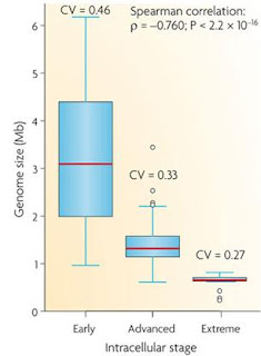
Correlation between genome size and the stage of adaptation to an intracellular lifestyle

Although genome size reduction is the overall fate of bacteria that form a partnership with eukaryotic cells, the process is more than simply the slow loss of genes over time. Different evolutionary pressures will act on both bacteria and host organisms at different stages in the process of creating a relationship which in the early stages requires the gain of genes allowing host interaction. In later stages, the bacterium will lose genes, in some cases passing them on to the safer environment of the host nucleus, which in turn can lead to the bacteria losing all its personal autonomy and becoming a eukaryotic organelle.


---
Toft C, & Andersson SG (2010). Evolutionary microbial genomics: insights into bacterial host adaptation. Nature reviews. Genetics, 11 (7), 465-475 PMID: 20517341

Graduation!

Finally finished! After four years of stupidly intense work and crazily long holidays I've finally officially finished my degree and left my university. I'm still living in the city, and odds are I'll come back for a PhD the year after next so it wasn't exactly a tearful goodbye, but I still managed to have fairly intense Moment when I scurried out the fire exit after having gone through the requisite amount of Latin for me to walk off with the actual degree.

I can now put letters after my name!

I've got around two weeks off before I start my summer project, which I am VERY much looking forward too. What with revision and the way the course works I haven't actually been in a lab since around March, which is far too long for a Lab Rat to be out of a lab for.

I also won't technically be a "lab rat" any more. I chose the name to mean "unpaid student lab worker" (it seemed vaguely funny at the time...) and now I am a fully fledged paid scientist (albeit one who is frantically looking for a job).

There will be more bacteria-related posts coming very shortly. This is just an apology-for-lack-of-science post because what with rehearsing for graduation, getting graduated, getting rascally drunk at graduation parties and looking for wedding dresses there hasn't been all that much time for bacteria.

~Lab Rat MSci(Hons) BA CANTAB :D

Bacterial Hitchhikers

ResearchBlogging.orgThere was an interesting post over at Culturing Science about the widespread dispersal of bacteria which, as well as sporting an amazing hand-drawn MS Paint picture also put forward the argument that bacterial evolution occurs in very selective environmental pockets and niches, while a sort of general 'less-evolving' population floats around the world. This helps to explain why you can find almost identical species of bacteria all over the world, yet still find very specialised colonies in distinct environmental niches. This can all be summed up with a rather nice little quote by the Dutch biologist Lourens Baas-Becking:

"Everything is everywhere; the environment selects"

It's a quote that can be hotly debated, but it certainly is true that bacteria have a remarkable capacity for spreading around the world. Even before humans and air-travel allowed pathogenic bacteria to go on holiday environmental bacteria have been spreading through the sea, air and soil not by their own (rather poor) motile ability but by hitching rides on other organism. In the case of soil bacteria they can also travel in spore form, which significantly reduces the harm they sustain by (e.g) being trampled all over the savannah's by an elephant.

In the sea bacteria have even more help with movement. Currents, tides, waves and general water movement can help to move bacteria large distances horizontally and a recent paper (that Lucas sent me, reference is below) provides evidence that bacteria might move vertically in the oceans by taking a ride on zooplankton, small eukaryotic ocean-dwelling creatures, a selection of which are shown below:

This isn't just a careless picking up of bacteria by idly floating zooplankton though, this is the bacteria actively attaching and dissociating from the zooplankton as they move through the water. The paper proposes a "conveyor belt hypothesis" which states that bacteria attach at one level, travel either upwards or downwards on the migrating zooplankton, and then dissociate when they reach where they want to be.

The reason bacteria would want to travel around between the different depths is due to nutrient availability (this may also act as a biochemical signal for the bacteria to fall off their zooplankton transporters). Deeper waters have a higher concentration of inorganic nutrients, while waters closer to the surface contain a higher concentration of oxygen, and algal derived organic matter.

In order to estimate how much travelling the bacteria were doing, the researchers used. Three different bacteria (that were thought to travel between layers, rather than bacteria that have adapted to the layer they are in) were isolated and labelled with GFP - a protein which fluoresces green. They were then added too migration columns, filled with zooplankton called daphnids which are very phototaxic (i.e they move towards light). Running a light up and down the migration columns lead to the daphnids moving up and down, and the movement of the bacteria could be tracked by following the spots of green florescence.

Sure enough, they found that the green bacterial dots would gather either at the bottom or the top of the migration column, but only when the daphnids were added (a column containing just bacteria and water with a light running up and down the side produced no results). They also found that the more Daphnids they added, the move bacterial movement was found, strongly supporting their hypothesis that the bacteria were taking a ride on the far larger zooplankton.
Graph showing the number of bacteria in the upper layer of the migration column per migration cycle of the dahpnia (i.e one movement up and down - 2 hours). The circle shows columns with no daphnia added, while triangle and square show 20 and 80 daphnia respectively. Numbers to the right are the regression slope.

Among other things, this study shows just how dependant ocean species can be on the other organisms they live amongst. The bacteria which use zooplankton for conveyor-belt style hitchhiking now have their own survival intrinsically linked with the continued well-being of the zooplankton species that they rely on. This knowledge can then be added to models of how large-scale changes to the oceanic environment will affect the creatures within it and ultimately, given the importance of zooplankton on the foodchain and the importance of oceanic bacteria on the environment, the fate of many other organisms.

---

Grossart HP, Dziallas C, Leunert F, & Tang KW (2010). Bacteria dispersal by hitchhiking on zooplankton. Proceedings of the National Academy of Sciences of the United States of America PMID: 20547852

---

Follow me on Twitter!

Archaea, Eukaryotes and the evolution of DNA replication complexes

ResearchBlogging.orgThe relationship between bacteria, archaea and eukaryotes is an interesting one, and made slightly harder to approach as people tend to lump archaea and bacteria into the one grouping of 'prokaryotes' which is not much more than a scientific word for "blobs I don't care about". Delving deeper into the biochemistry of all three superkingdoms shows that while the metabolic pathways used by archaea are more similar to those in bacteria, their core DNA processes (such as replication and protein synthesis) are more similar to the processes in eukaryotes. (I talk more about the distinction between the three superkingdoms here)

There was an interesting paper in PLoS ONE lately that was looking at the evolution of DNA replication complexes in archaea, and seeing as this blog has been rather heavily bacteria-biased (i.e I haven't talked about archaea for a while) I decided to take a look at it. They were focussing on three main complexes that help in DNA replication and are found in both archaea and eukaryotes: proliferating cell nuclear antigen (PCNA), replication factor C (RFC), and the minichromosome maintenance (MCM) complex. Bacteria do use corresponding proteins, but they are far more distantly related.

Schematic of the structure and subunits of the three complexes.

The MCM complex is thought to act as a helicase; unwinding the two DNA strands to allow them to split in two to be replicated. The PCNA and RCF are known as the clamp and clamp loader and help to attach the RNA primer for replication to the Polymerase, which uses the primer to start replicating the DNA.

All of these three complexes consist of separate subunits, which are almost identical. PCNA, for example, is a trimer (in the diagram above each subunit is a separate colour). In eukaryotes these the subunits are identical, but in archaea variations are found between them. This general pattern, that subunit composition was far more variable within the archaea, was found in all three of the complexes. This method of gene duplication followed by gene modification to create two different proteins is an important one for evolution, and in the case of DNA replication it seems to have been exploited far more in archaea than in eukaryotes.

Changing some subunits also allows these complexes to carry out different tasks. It's been suggested that for some archaea there may be a functional difference between PCNA with all subunits the same (homotrimers) and PCAN with differing subunits (a heterotrimer). This allows multiple functions to be generated through simple DNA duplications - although all the functions are likely to relate to DNA replication in some way.

This brings forth the interesting point of view that the truly 'ancestral' forms of these genes and proteins may be more like the proteins seen in the eukaryotes rather than the archaea! Archaea (and bacteria) can tolerate a lot more genetic change than eukaryotes can, and have a far shorter generation time, allowing them to change and evolve more quickly than the larger, less genetically mutable eukaryotes. On the other hand the lack of change and high level of conservation in eukaryotes means that the complexes remain very similar to those of the ancestral eukaryote from which they evolved. They may even be closer to the forms found in the last common ancestor between eukaryotes and archaea, before the eukaryotes gained a nucleus and became unable to share genes with the surrounding organisms.

---

Chia N, Cann I, & Olsen GJ (2010). Evolution of DNA replication protein complexes in eukaryotes and Archaea. PloS one, 5 (6) PMID: 20532250

---

Follow me on twitter!

Colony behaviour and metatranscriptomics

ResearchBlogging.orgMost places which contain bacteria tend to contain lots of them. In the environment (i.e outside human bodies) bacteria often live in large colonies which can make it difficult to explore their reactions to changing conditions. In the lab, with just one bacteria, information about responses can be obtained by transcriptomics; looking at how the transcriptome changes as the environment does.

The transcriptome is the set of all the mRNA within the cell. Unlike the genome, which is the all DNA present within the cell, the transcriptome only reveals those genes which are being turned into proteins. This therefore acts as an indication of the changes in protein production within the cell.

My MSPaint skills are improving.

For large bacterial colonies however, individual transcriptomic studies aren't much use for finding out the state of the whole colony. Each bacteria within the colony will not only be reacting slightly differently, but will also be experiencing different conditions within the colony depending on where it is within the colony. It's often best, therefore to treat the entire colony as one 'cell' and carry out transcriptome studies on the whole lot. This is metatranscriptomics.

Nowadays one of the easiest ways to carry this out is by isolating the RNA and getting it all directly sequenced by Pyrosequencing (which saves the trouble of making microarrays). As well as giving information about the changes within a colony due to environmental factors, it can also show the changes in protein production at different stages in the colony lifecycle such as at the beginning and end of an algal bloom. The integration of new third-generation sequencing methods into this process will make it faster and hopefully allow isolation of the rarer, less abundant transcripts to find more subtle changes in gene expression.

This has important implications for things like oceanic cyanobacteria which are involved in carbon sequestration in the oceans. Understanding how changes such as increases in ocean acidity (or decreases in salinity) affects their growth and ability to remove carbon from the atmosphere could have important implications for global warming, and how it can be dealt with.

---


Gilbert JA, Field D, Huang Y, Edwards R, Li W, Gilna P, & Joint I (2008). Detection of large numbers of novel sequences in the metatranscriptomes of complex marine microbial communities. PloS one, 3 (8) PMID: 18725995

Guest post

I've written a post for Lucas at Thoughtomics about how some bacteria can act as little compasses which you can find here. Lucas's blog has some really amazing posts, and he was a biology finalist for the researchblogging.org awards so I would recommend checking out some of the stuff he's written as well.

He also wrote a guest post for my blog a while ago (which you can find here) so this is me returning the favour.

Should be a post on oceanic bacterial metatranscriptomics coming up next week!

Moving house...

The more perceptive of my regular readers may have noticed that my blog has now moved to Field of Science, which gives me a snazzy new URL and the chance to be among some amazing science bloggers including blogging-friends like Psi. As I mentioned, this blog will now be kept strictly prokaryotic, providing (hopefully) interesting information about all things bacterial and archaeal. I usually update twice a week, three times a week if I'm feeling particularly enthusiastic, with occasional brief holiday breaks if I'm away somewhere without internet access (although it's not looking like I'll be having any of those for a good few months now...)

One of the main things I don't do on my blog is mention personal details. This blog is kept strictly for science and almost exclusively for research blogging, with the occasional bit of scientific personal speculation. However I do have some rather awesome breaking news...

I just got my Finals results - 2:1 !

If I can't show off about that on my own blog, where can I show it off?

There won't be a post today as I'll be celebrating by watching back-to-back "Auf Wiedersehen, Pet" episodes and waiting for my partner to come home from the late shift, but I've got one coming up tomorrow about the trials and tribulations of bacterial intracellular parasites.

Strategies of Intracellular Parasites

This post was chosen as an Editor's Selection for ResearchBlogging.orgIntracellular parasites have a difficult life. On the one hand, they need to utilise the resources of the host cell, which will ultimately cause it damage, however on the other they must necessarily keep the host cell alive so that they can live off it. This is particularly difficult when your host cell is a eukaryote cell in a multicellular organism, because multicellular organisms can't afford to have one cell behaving oddly. If the cell starts to notice any differences in its behaviour, it promptly commits suicide, meaning that in order for the parasite to survive it has to find ways of preventing the cell from killing itself when it notices things going wrong.

Chlamydia is a particularly well studied human intracellular bacteria, and is best known for being sexually transmitted and featuring in posters on student welfare notice boards in probably every university in the UK:

If it's worth talking about, it's worth blogging about!

The reason Chlamydia is so worth talking about is two-fold, firstly because it's a bacterium rather than a virus and can therefore be (relatively) easily treated with antibiotics. Secondly, because you don't always know you have it and therefore it's worth getting tested even if you don't appear to have any symptoms. The bacteria can live perfectly happily within your cells replicating away without the body noticing, and if left untreated can lead to quite serious problems, such as blindness, pelvic inflammation or sterility.

In order to remain replicating inside the cells unnoticed for so long, Chlamydia have to prevent the cells from either destroying them or committing suicide before they manage to replicate. One of the ways they can do this is by degrading the cell proteins involved in cell signalling pathways. An example is the serine protease (i.e an enzyme that breaks down proteins) CPAF which is secreted by the infective chlamydia particles and (among other things) breaks down the protein HIF-1 which is used to trigger the cell suicide response to low oxygen levels. They can also break down proteins which would potentially be involved in the immune responses to the damaged cell, such as NF-KB, which helps activate the innate immune system inflammatory response.

Quite how the chlamydia causes the host cell protein degradation is still a little unclear. They may use the common viral strategy of modifying proteins to make them more susceptible to being picked up by the cellular degradation machinery (although there is no biochemical evidence for this as yet) or alternatively it might activate protein degradation pathways that are usually silenced in uninfected cells. Analysis of the chlamydia genome shows several predicted proteases, so it may be possible that rather than using host proteases the bacteria is degrading specific proteins with its own protease enzymes. The bacterial protease CPAF (mentioned above) has been crystallised and the crystal structure shows the potential for several different substrates (i.e it could potentially degrade many different proteins) so it might play in important part in this process.

The use of degradation proteins also creates a potential target for therapeutics. If that sentence sounds familiar it's because I've written it several times before and will probably write it several times in the future. "New targets for therapeutics" is pretty much THE standard excuse for studying anything related to bacteria. From a more selfish and less funding-motivated standpoint, it also provides exciting new information about how bacterial and eukaryotic cells interact, and how parasites that live inside cells can control host signalling pathways to their own advantage.


---

Zhong G (2009). Killing me softly: chlamydial use of proteolysis for evading host defenses. Trends in microbiology, 17 (10), 467-74 PMID: 19765998

---

Follow me on Twitter!

Antibiotics and Synthetic Biology

ResearchBlogging.org
The model for bacterial death by antibiotics was fairly simply until recently. Antibiotics work by targeting a certain area of the bacteria; beta-lactams target the cell wall, Rifamycins target RNA synthesis, tetracyclins inhibit protein synthesis etc. The theory was that by inhibiting these processes, a certain vital function within the bacteria would be stopped, leading to its death.

However due to research done by Kohanski (references below) the story is looking a bit more complicated. Looking at three different classes of antibiotics they found that no matter what the site of action, all the antibiotics induced hydroxyl radicals. This was in bactericidal drugs, which actually kill bacteria, rather than bacteristatic ones (which just prevent cell growth). They also demonstrated that this mechanism of hydroxyl radical production was the end product of a chain of reactions involving damage to the TCA cycle (aka the Krebs cycle - which is a major part of respiration) which lead to damage to iron-sulphur clusters and subsequent production of the DNA-damaging hydroxyl radicals. This is shown diagramatically below, and this first paper was covered by Jim at Mental Indigestion with some great follow-up comments and discussion.


They've recently put out a review (second reference below) of which I find the most exciting parts are the two little extra-information boxes. One of them covers drug synergy and the second covers synthetic biology, both of which I'm getting increasingly more interested in.

Drug synergy

One of the most useful things about modelling drug actions is it can help to show which drugs would work most effectively in pairs. Using two drugs together can have many potential effects; it can make the treatment more effective, sometimes is can make the treatment less effective and of course some can be dangerous for the patient. Work on drug synergy showed that aminoglycoside antibiotics (which affect RNA synthesis) become more affective when given simultaneously with B-lactam antibiotics (which lead to cell wall breakdown) as the increased cell wall breakdown helps the aminoglycosides to get inside the cell. Conversely, drugs that inhibit protein synthesis are less effective when given at the same time as drugs which inhibit DNA synthesis as making it harder to synthesise proteins from sub-optimal DNA actually makes the cell more able to survive.

These interactions will affect the dosage of drugs used during synergistic treatments, and it is hoped that using two different types of antibiotics at low doses might be more healthy for the patient, and might help to combat against antibacterial resistance to one of the drugs.

Synthetic Biology

Another interesting concept the paper brings attention too is the potential use of synthetic biology to aid in both the study and application of antibiotic-related death systems. By using synthetic genes to disrupt or alter the proposed antibiotic network novel drug targets could be discovered. If turned into a high-throughput system this would be far more useful than the current screening system which tests for a potential drugs interaction with a target, rather than the ability of this interaction to lead to cell death.

Synthetic genes can be delivered into the bacterial cell via bacteriophages. Adding a synthetic gene into a bacteriophage for bacteria cell delivery has been attempted successfully before when they were used to enhance E. coli cell death by delivering genes for proteins that disrupted the DNA-repair system within the bacteria. This allowed faster and more effective killing of the bacteria at lower doses of antibiotic.

At a time when bacteria are fast becoming resistant to even the front line drugs, research that suggests novel ways of killing bacteria can produce some very useful outcomes. Using combinations of drugs at lower concentrations, or aiding antibiotics by introducing them along with synthetic genes in bacteriophages allows an increased shelf-life of the drugs that we currently possess as well as providing potential systems to aid the discovery of new antibiotics.


---

Kohanski MA, Dwyer DJ, Hayete B, Lawrence CA, & Collins JJ (2007). A common mechanism of cellular death induced by bactericidal antibiotics. Cell, 130 (5), 797-810 PMID: 17803904

Kohanski MA, Dwyer DJ, & Collins JJ (2010). How antibiotics kill bacteria: from targets to networks. Nature reviews. Microbiology, 8 (6), 423-35 PMID: 20440275

---

Follow me on Twitter!

Niches of Sunlight

ResearchBlogging.orgIn every environment there will be competition for resources, and there are generally two ways organisms deal with this; generalise or specialise. To generalise, you try to cope with as many different conditions as possible, so that if you get out-competed in one area you can try to cope with the conditions elsewhere. To specialise, you get damn good at using the conditions in your little niche, in the hope that you'll be better than anyone else who comes along, and be able to out-compete them.

There are many resources that need fighting over, and in the sea one of the major ones for photosynthetic organisms is sunlight. Other nutrients such as nitrogen, phospherous and trace metals such as iron and copper can exist in different forms at different levels in the ocean (as shown below), but once you start getting below a certain depth, sunlight quickly becomes a finite and rapidly diminishing resource:
Diagram showing availability of nutrients at different depths.
Taken from the reference below.

Different organisms can cope with the lack of sunlight in different ways. Some (especially the larger algae species) have generalised, they contain a whole range of different light capturing pigments which can absorb a range of light wavelengths, including those in the darker depths. But little photosynthesising bacteria like Procholorococcus (which I mentioned in this post) which have the smallest genomes of all photosynthesising organisms, don't have that option. Instead they have to specialise, so that different strains in a species are adapted to different levels of light.

Work done by Rocap (paper reference below) looked at two different strains of Prochlorococcus: MED4 and MIT9313 (which I will just call MED and MIT). The MED strain was found only in the surface waters, while MIT was found much lower down; a phenomenon known as 'vertical niche partitioning'. Despite their genomes differing by only 3%, and despite being technically the same species (although 'species' is an uncertain word in the world of bacteria) they have optimised themselves to completely different levels of not just light but also nutrients, trace metals and virus specificities.

MED (the one near the surface) has a slightly smaller genome than MIT, yet contains twice as many genes dedicated to high-light-inducible proteins, many of which seem to have arisen by gene duplication. It also has genes specialised for the nitrogen sources found near the surface of the water, and organic phosphates (which again are found predominantly on the surface).

MIT on the other hand has fewer genes for ultraviolet damage repair, but more light harvesting genes, for example it possesses two copies of the hight-harvesting chlorophyll binding antenna protein. This helps it to gather as much light as possible, despite being further below the surface. it's also adapted for its specific nitrogen source and increased ability to use orthophosphate, rather than organic phosphates.

Both genomes have lost the ability for photoacclimatisation, that is the ability to change to suit different light conditions. By taking up vertical niche positions, they have forfeited the ability to change their response, meaning that a strict horizontal partition between them must be maintained at all times. Any Prochlorocuccus found at lower levels will be of the MIT variety, while those at higher levels will be MED. It's even thought that there might be further strict niche partitioning; with different ecotypes of MED adapted to use different iron sources, or different temperatures.

For the photosynthetic organisms that inhabit it, seawater is more than just a blue shifting salty mass. It's a whole range of niches and environments, partitioned in three dimensions depending on the surrounding conditions and nutrients.

---

Rocap G, Larimer FW, Lamerdin J, Malfatti S, Chain P, Ahlgren NA, Arellano A, Coleman M, Hauser L, Hess WR, Johnson ZI, Land M, Lindell D, Post AF, Regala W, Shah M, Shaw SL, Steglich C, Sullivan MB, Ting CS, Tolonen A, Webb EA, Zinser ER, & Chisholm SW (2003). Genome divergence in two Prochlorococcus ecotypes reflects oceanic niche differentiation. Nature, 424 (6952), 1042-7 PMID: 12917642

---

Follow me on Twitter!