Field of Science

Bacterial comet tails

ResearchBlogging.orgI haven't worked very much with bacteria that infect humans. Most of my lab work has been done in the fields of either synthetic biology (which works with model organisms) or antibiotic production, which works on soil bacteria that produce the antibiotics. Human bacterial parasites therefore hold the fascination of the slightly exotic, not least because they sometimes do things like this:

Figure from"Molecular Biology of the Cell, Fourth Edition"by Alberts et al.

I've written before about some of the interesting features of intracellular bacteria, but this is possible one of the more exciting and fun things that they do. The picture above shows a eukaryote (human) cell outlined as a green oblong. Within the cell are lots of invasive bacteria (the red dots) some of which have a beautifully long green 'comet tail' flying out behind them.

That comet tail isn't just for show, it is vitally important for movement. The inside of a eukaryote cell is a fairly crowded and busy place, bacteria can't just swim around inside the cell like they would in the wild. Instead they have to rely on physical methods to push them through the cell and like invading virus's (which I wrote about here) they hijack machinery inside the cell to move them around.

Virus particles can latch onto the intracellular transportation machinery to hitch a free ride, but bacteria are too big for that. Instead, what most of them do is to produce proteins known as nucleation-promoting factors. These co-opt cellular proteins (the Arp2/3 complex for anyone with a background in actin polymerisation) which form branched actin fibres behind the bacterial cell, pushing it forward. The 'comet tail' pattern seem above, is seen by using a green stain for the structural actin protein, so you can see it forming long fibrous complexes behind the bacteria. These actin tails can move the bacteria wherever they want to go in the cell, and can also help with the invasion of neighbouring cells. This process is shown diagrammatically below:

Figure from"Molecular Biology of the Cell, Fourth Edition"by Alberts et al.

The actin system is a good one to use, as actin is ubiquitous inside all eukaryotic cells. In normal cell conditions is it used for structural purposes and is vital in cell division. Also important for the pathogenic bacteria (which after all does not want to kill its host straight away) is that none of these important cellular processes are compromised by the bacteria 'borrowing' some of the actin to move around with.

Another interesting point is that different intracellular bacteria often produce different types of actin tails. L. monocytogenes and S. flexneri have short, highly crosslinked filaments producing short stubby little tails, whereas Rickettsia species have actin tails that are composed of distinctly longer bundles of unbranched actin filaments. Part of the reason for this is that different bacteria will produce different nucleation-promoting factors and some of the more lazy ones (i.e S. flexneri) don't even bother to do that and just use the host nucleation-promoting factors within the invaded cell! Recent work has shown that Rickettsia on the other hand, doesn't even rely on the host Arp2/3 complex to polymerase the actin and instead relies almost entirely on their own, bacterial, proteins.

They truly are beautiful to look at though. Even without all the fancy colour staining:


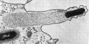
Listeria monocytogenes pushing right at the cell membrane, with actin tail behind. Electron micrograph picture taken from the se reference below

---
Ray K, Marteyn B, Sansonetti PJ, & Tang CM (2009). Life on the inside: the intracellular lifestyle of cytosolic bacteria. Nature reviews. Microbiology, 7 (5), 333-40 PMID: 19369949

Kuo SC, & McGrath JL (2000). Steps and fluctuations of Listeria monocytogenes during actin-based motility. Nature, 407 (6807), 1026-9 PMID: 11069185
---
Follow me on Twitter!

Oil-eating bacteria

ResearchBlogging.orgOil is formed from hydrocarbons: organic compounds which consist soley of the elements hydrogen and carbon. There are many, many different types of hydrocarbons, all of varying lengths and shapes, and pretty much all of them can in some way be consumed as an energy source by bacteria.


Hydrocarbons, in both ring and chain form, taken from a mix of sources

The general rule is that the shorter and fewer rings present, the more toxic the compound to bacteria (ethanol, for example, is deadly) however there are very few hydrocarbons that some bacteria somewhere won't consume. Like all living organisms, bacteria need a carbon source for energy, and when there are none of the coventional ones around (such as glucose and lactose) many of them will start consuming the ones in oil - crude oil contains a lot of carbon, which is why we burn it for energy in the first place.

Oil-eating bacteria have been found in a huge number of locations as well - seawater, freshwater, groundwater, water containing sludge, and land environments such as silt, soil and sand. They are found in arid deserts o the Middle East (which to be fair is a good place to hang out if you consume oil) and even in ice cores from Antarctica. And rather than being found widespread within seperate species, the capacity for oil consupmtion is often found in many different species within a particular location - in the bacterial world you are more likely to share habits with your nearest neighbours than with your closest relatives.

Using these bacteria for our own purposes, however, is proving slightly more problematic. You can purchse freeze-dried hydrocarbon-degrading bacteria and sprinkle them on your oil slick like minature Captain Planets, but their abilities are currently not nearly as effective as chemical detergents, or physical removal of the oil. This is mostly because these freeze-dried bacteria are specialised lab strains that (much like several of the academics that constructed them...) are often highly unsuited to the real world outside of the safe confines of the lab. Sometimes as well the pollutant is unaccessible to the bacteria, which may be floating on the top of the water while the oil is in droplets underneath the surface.

Looking into the potential of oil-eating bacteria is encouraging though, especially after incidents like the BP oil spill earlier this year. For the oil eating bactera that were hanging around, that must have been a feast day! And even though that event has now been largely forgotten by the media, there will still be coils and dropplets of oil floating around from it, and those will be cleaned up almost exclusively by bacteria.

---

Lena Ciric (2010). A natural solution: how bacterial communities can help clean up oil spills Microbiology Today, 229-231

---
Follow me on Twitter!

Breaking Biofilms with DNA

ResearchBlogging.org I've written about biofilms a couple of times before but it's an interesting enough topic to keep returning to. As a brief summery, biofilms are large collected colonies of bacteria, often surrounded by a sticky mesh of glycoproteins. They are ultra-annoying in the case of infectious bacteria as the bacteria deep in the depths of the biofilm will not be exposed to any antibiotics, the layers of glycoprotein and surrounding bacteria will protect them.

Although living within a biofilm contains significant advantages (protection, good living conditions, etc) there are also times when the bacteria will want to swim away, in order to disperse and form new colonies. The bacteria C. crescentus has an interesting way of doing this, each round of cell division produces two cells: a moving 'swarmer' cell and a non-motile 'stalked' cell which attaches to the biofilm, or any other surface. If conditions are right, the swarmer cells swarmer cells will eventually turn into stalked cells; loosing their flagellum (which are used to swim), retracting their pili, and growing a membranous 'stalk' to attach it to surfaces or surrounding bacteria. This is shown below:


Lifecycle of Caulobacter crescentus - image from reference 1

Exactly what it was that turned the motile cells into stable ones and maintained the biofilm was not well understood. Recent research found, rather excitingly, that one factor that could lead to the maintenance of swarmer cells, and the breaking up of biofilms, was extracellular DNA (eDNA) - i.e genetic material that had escaped from cells and was floating around the biofilm. Adding eDNA to C. crescentus biofilms lead to biofilm dispersal, an affect that was reversed by adding DNase enzymes that broke down the DNA.

Why is eDNA such an important signal? Because it's one of the most common products produced from dying cells within a biofilm. Once bacteria in a biofilm die, their cellular integrity breaks down, their insides become their outsides and their genetic material spills out into the surrounding area. This can then act as a powerful signal for surrounding cells, and if the cells around you are dying then where you currently are is clearly not a good place to be. This is also useful when areas of biofilm start to get saturated with too many bacteria, just a few dying off will clear the way for new swarmers to leave and maybe set up colonies elsewhere.

---

Berne C, Kysela DT, & Brun YV (2010). A bacterial extracellular DNA inhibits settling of motile progeny cells within a biofilm. Molecular microbiology PMID: 20598083

Jermy A (2010). eDNA limits biofilm attachment. Nature reviews. Microbiology, 8 (9) PMID: 20737663

---
Follow me on Twitter!

Antibodies

I'm away this weekend sorting out weddings and grad applications, so have a video! This is probably the best explanation of antibody structure I've ever come across, so well worth watching for anyone trying to wrap their head around the immune system. As a bit of background information antibodies are what bind to antigens (i.e molecules that are recognised as not being part of the body and may be pathogenic) and can help destroy them. Antibodies must therefore have a very strong and stable structure, but also must have very variable regions that allow different antibodies to recognise a range of different antigens.


Essential Cell Biology, Second Edition
by Alberts, Bray, Hopkin, Johnson, Lewis, Raff, Roberts, Walter
copyright 2004 by Garland Science Publishing

And i'm still looking for more posts for the MolBio carnival on the 6th December! If you've written anything concerning the innards of cells over the last month, please submit your post here - carnivals are a great way of gaining readers and getting yourself noticed.

Storing DNA

ResearchBlogging.orgDNA is one of the most important components of the cell. In eukaryote cells (i.e the cells of humans and plants) it is stored inside a nucleus that keeps it safe and away from dangerous things like free radicals produced by the metabolic reactions of the cell. In bacterial cells the DNA isn't nearly as well protected, but the main bulk of the bacterial chromosome (excluding the little floating plasmids) is all kept together in a bundle usually referred to as a nucleoid.

However the DNA in cells is rather long which means that in order to package it into a small space it needs to be coiled up. Eukaryotic cells do this by using proteins called Histones, which coil the DNA around them, and can also signal which genes the cell should be turning into proteins.

Image from wikimedia

Because eukaryotes cells have so much DNA they need to keep it all tightly coiled up, with little molecular tags on the histones to remind them which bits are needed reguarly, and which bits can be mostly ignored. Prokaryotes on the the other hand don't need quite as much control, most of their DNA is going to be used most of the time, but it still needs to be packaged up to fit inside the cell.

The cells do this using by using the unbelieveably eukaryot-ist named "histone-like proteins" such as H-NS which is found in E. coli and related bacteria. Although their precise mechanisms are not quite clear, they have been shown to play a clear role in the coiling of the DNA and maintaining nuceolide stability. They also (like their eukaryote counterpart) help to control gene expression as well as chromatin structure.

In most cases, the H-NS functions as a transcription repressor merely through its physical presence wrapped around DNA - it's harder to transcribe DNA (the first step for protein production) when it's all wrapped up tightly in balls. Some genes though, seem to be activated when associated with H-NS, as it can affect the stability of the mRNA transcript. These are usually genes associated with stress responses; in fact many of the genes that are affected by H-NS are linked to the stress response or changes in the environmental conditions, such as high or low temperature, high osmolarity, changes in pH or oxygen concentration.

Quite how the H-NS controls these parts of the DNA is not entirely clear. Unlike histones, which are positively charged to stabilise the negative charge on the DNA, H-NS proteins are neutral. Although the domain of the molecule that binds to the DNA has been identified, it is not yet certain which parts of these are vital, or how they interact with the DNA molecule. One thing that is clear however is that H-NS are capible of forming dimers, and probably carry out much of their task as a dimer of two joined molecules.

Domains of an H-NS molecule, from the reference. The oligomerization domain binds to a fellow H-NS while the DNA binding domain attaches to DNA.

Whatever the binding mechanism is, it is unrelated to the actual sequence of the DNA, as H-NS can bind to many different regions of the gene, regardless of sequence. Until recently the H-NS molecules were thought to have no post-translational modifications (unlike histones which are often decorated in molecular markers to indicate which kind of gene they are on) but some have recently been found to be marked with poly-3-hydroxybutyrate, a small lipid molecule. The reason for this is unclear, but it does raise some exciting implications for H-NS control of gene transcription.

When it comes to controling DNA expression, it's clear that in both eukaryotes and prokaryotes, the scaffold proteins that hold the DNA coiled up act as more than just a scaffold. Instead they are involved in a substantial amount of the control of DNA expression, often working closely with other control proteins to ensure the correct genes are turned into proteins.

---

Schröder O, & Wagner R (2002). The bacterial regulatory protein H-NS--a versatile modulator of nucleic acid structures. Biological chemistry, 383 (6), 945-60 PMID: 12222684

---
Follow me on Twitter!

Exciting Things

A couple of fairly exciting things have happened in my internet-world over the last few days and I thought I'd share them:

I HAVE A GUEST BLOG POST AT SCIENTIFIC AMERICAN! I am really excited about this as it gave me a chance to write a proper essay-style blog post with a good load of references. It's also a good place to make your point of view known, so if you have anything you've ever wanted to say about synthetic biology, or the ethics and applications of, go stick a note in the comments - here. I'd relaly appreciate it and so would BoraZ :p

Huge amounts of thanks must go to Bora Zivkovic (twitter @BoraZ and blog on the link above, who gave me the chance to write there.

iGEM!
I am really proud of the Cambridge iGEM team, who made the finalists this year, and also won an award for best wiki (read it here!).
There was a lot of love for the Gibson Assembly song as well. But in general I think an amazing amount of effort has gone into this from all the iGEM teams, I hope they all had an amazing time and enjoyed the conference in Boston. The full results of the competition can be found here.

CARNIVAL TIME! The next edition of the MolBio carnival will be held here at Lab Rat and I'd love to make it a nice big one. If you've written any MolBio articles this month, or plan on writing any, please submit them all here - and I'm usually fairly easy on the strict definition of 'molbio' anything fairly small and scientific will do! If you know anyone who writes good molecular biology posts, go and pester them to submit as well - carnivals are a great way to increase the readership of your blog, and to get your name spread around the internet a bit.

Things are also quite exciting in the real world as well, what with wedding plans, PhD applications, job searching and the first positive-ish results I've had in the lab for a while. Life in general is quite exciting for me at the moment (and to be honest, I think it always has been :D )

Antibiotics and gut bacteria

This post was chosen as an Editor's Selection for ResearchBlogging.org
All microbiologists end up writing about gut bacteria at some point. It is the way of things. Disease of the Week is currently doing a whole series on it, and a few weeks ago I covered the interaction of the immune system with gut bacteria (here). However a recent paper came out in Microbiology Today concerning the affect of antibiotics on gut bacteria, which is a topic that I both find interesting and have had some actual experience with.

I've taken antibiotics a few times, and each time I've found that despite the many positive effects it has (i.e I don't die of septicaemia) being on antibiotics tends to make my stomach fairly unhappy. I usually have a good relationship with my stomach - I feed it regularly with plenty of food and in turn it is generally quite accepting of the fact that I don't always wash my hands as much as I should, and occasionally eat things that might be past their sell-by date, or heated up food that isn't exactly the "piping hot" recommended by the label.

Antibiotics however are usually designed to kill off bacteria, and unless the antibiotic in question is very specific that often includes your commensal gut bacteria; the 'friendly' bacteria that yoghurt companies keep mentioning, that hangs around in your gut and prevents other bacteria invading. In the short term this leads too the occasional unhappy stomach but in the long term it can have a more sinister effect, by encouraging antibiotic resistance genes to develop and spread amongst the normal bacteria in your gut.

The diagram below shows a stylised (and not to scale) diagram of what happens to your gut bacteria after a dose of antibiotics. The antibiotic resistant bacteria (in purple) are suddenly at an advantage and can proliferate. As time goes by, the other natural bacteria gradually return to your gut (which is not after all the most sterile of environments) but there will be a higher concentration of antibiotic-resistant bacteria around.

Diagram taken from the reference (below)

It's important to remember that these antibiotic-bacteria are not dangerous in themselves. Unless they move out of the gut for any reason they will remain totally harmless. However they also contain the genes for antibiotic resistance, and if another bacteria manages to survive and get into the gut, they can pass those genes on. As every human carries different gut bacteria, and will take different antibiotics in the course of their lifetime, this study cannot really make any hard and fast rules about what the effect of this might be on any one individual, but it does make the point that many people will be carrying within them a large proportion of bacteria that may have developed a novel form of antibiotic resistance.

It's not always terrible, in some cases the antibiotic resistant bacteria are grossly unfit, and are immediately out-competed as soon as the antibiotic goes away. Those that do survive both the antibiotic and the returning bacteria however, can hang around for a long time, and antibiotic resistance was seen in gut bacteria up to four years after antibiotic treatment had finished. And the intestines are such a wonderful environment for bacteria to share DNA in; there are abundant nutrients, everything is all moist and warm and there are a lot of bacteria in very close contact with each other.

If you have taken antibiotics it's not worth loosing sleep over, but it's something hospitals are starting to be more and more aware of.

---


Jernberg C, Löfmark S, Edlund C, & Jansson JK (2010). Long-term impacts of antibiotic exposure on the human intestinal microbiota. Microbiology (Reading, England), 156 (Pt 11), 3216-23 PMID: 20705661

Waking sleeping bacteria

ResearchBlogging.orgThere are quite a few bacteria that, when times are hard, are able to put themselves into a state of metabolic inactivity. They are still technically alive - just not really eating, growing or changing in any way, just waiting until conditions become more favourable so that they can regain normal active behaviour. The bacteria used most frequently to explore this behaviour is B. subtilis which frequently form spores in response to adverse conditions, such as lack of nutrients or extremes of temperature.

Getting into a dormant state is fairly understandable, bacteria have lots of sensing systems which can tell whats going on in their surrounding environment, and dormancy is just a response to that. However getting out of a dormant state is a little more complicated. Once the cell is dormant hardly any of it is active, whatever signal system is used to activate the cells, it has to require very few cellular componants.

In some bacteria (particularly E. coli persisters) exit from dormancy seems to be a fairly random, stochastic effect. Every now and then, one of the cells will simply switch back to being active. If conditions are good it will replicate and re-colonise, whereas if conditions are bad it can either switch back to dormancy or, if it dies, be later replaced as its fellow bacteria come back. It's not a wonderfully good strategy for individuals, but for the species, and for the genes, it works very well.

One possibility the paper in the reference was looking at is that bacteria exit dormancy in response to growth signals from other surrounding bacteria. The idea behind this is that once bacteria start growing and dividing they start secreting signaling molecules such as muropeptides from the cell wall. If these muropeptides are present, it means that bacteria are able to grow and survive, and this might serve as a signal to other bacteria: "conditions are good enough for us!"


Diagram from the reference. a) bacteria switching from dormancy (light brown) to growth (green) in response to environmental signals. b) growing bacteria releasing signals to activate surrounding bacteria.

Addition of amino-acids and other required nutrients can activate B. subtilis spores, but the concentrations of amino-acids needed are so high that this is unlikely to be a natural response. On the other hand, addition of even small amounts of muropeptide leads to spore activation, strongly suggesting that it may be a relavent signal.

With this and the data from stochastic E. coli persisters it is tempting to see this as a full mechanism: exit from dormancy is random, but once one bacteria has grown and survived it can signal to the others that conditions are safe for growth. The only problem with this is that it has the potential to produce large amounts of bacterial growth independent of any signal from the actual environment. It has also been found (via "unpublished observations") that germinating spores don't release the same kind of murapeptide signal, and are unable to activate surrounding spores.

It was also found that the murapeptide signal was not really species specific, which is fascinating from the point of view of bacterial ecology. Growing bacteria, especially Gram-positive species, release large quantities of muropeptides as they grow, and it is a very well conserved molecule, found in the cell wall of other bacteria. A dormant bacteria might therefore not even need one of its own species to report back on conditions outside the cell, it can rely on signals from other bacteria in the surrounding environment.

---

Dworkin J, & Shah IM (2010). Exit from dormancy in microbial organisms. Nature reviews. Microbiology PMID: 20972452

---
Follow me on Twitter!

Interview with the Lab Rat

A while ago, I got sent an email with a short little list of questions asking whether I'd like to be interviewed for the Science and Technology section of the Charlotte Observer. To give an idea of what they wanted they sent along a couple of examples. I wrote out a couple of answers, sent it off, and was very surprised and excited when I found out today (via Bora's twitter) that the interview has actually been published! You can read it HERE.

The more perceptive among you may have noticed that included in the interview is a photo. The photo is of me. After spending two years being paranoid about the internet world connecting a picture of SE Gould (no relation to the Other Gould!) to the persona of Lab Rat, I've gone and sent over a nice big pic of my face for them.

I thought I'd better put forward a few reasons for this.

1 - The ego reason. They wouldn't publish the interview without a picture and I really wasn't about to pass up on an actual interview.

2 - The feminist reason. All the examples they sent me had pictures of men. I really wanted to get a picture of a woman in there somewhere.

3 - The 'I am fed up of being scared and paranoid' reason. This one takes more explanation. Suffice to say because of where I was brought up I was constantly surrounded by this "be doubly careful of everything you do and everyone you talk too" mindset. Even now, back in England, it's everywhere. Walking home in the dark, going travelling alone, being alone in a room with a stranger, I seem to be constantly slightly jumpy that something might hurt me. And all the while I am surrounded by people who are doing the same things as me and don't care about the danger at all. I know what almost all of my bloggy friends look like (with the exception of Psi); they aren't getting mass-murdered so why should I be scared?

But yeah, it was mostly the dazzling prospect of an interview that broke that principle :p

[And anyone who wants to take the picture and feature it in a "top ten sexiest microbiologists!" list: don't. Just don't. It'll piss me off, and will also be a good sign that you need glasses.]