Field of Science

Bacterial cell division and membrane potential

ResearchBlogging.orgBacterial cell division is usually quite a regular business. As I mentioned previously, not all bacteria use the regular FtsZ ring method of dividing, but for those that do division is mostly a matter of lining the right proteins along the middle of the bacteria, and then contracting a little ring of protein (FtsZ) around the centre of the bacteria to split the one cell into two cells.

Many of the more critical proteins in the process are membrane-bound, in particular the Min proteins, which in E. coli accumulate at the cells poles and prevent formation of the FtsZ ring anywhere but the centre of the cell:
A very simple diagram showing the Min proteins in fuzzy blue at the poles, and the FtsZ ring in red in the centre of the cell. This ring then contracts to create two bacterial cells.

So a group of researchers in Newcastle were working on the Min system and looking at it by immobilising the bacteria on slides which were covered in a layer of polylysine. They were finding it very difficult to actually get this localisation of the min proteins, rather than being membrane bound at the poles the protein was all over the place. They did a few more tries and eventually realised that it was the polylysine that was causing the problem. Adding polylysine to cells trying to divide slowed the process right down as it stopped the Min from properly localising.

What polylysine does to cells is affect the proton-motive force (pmf), which is used by bacteria to produce energy.

Diagram shows a stylised version of proton motive force at the bottom of the bacteria. Ions are pumped through the membrane creating energy in the form of ATP. Image taken from Osaka university website.

In order to verify that the crazy-behaving Min proteins were due to the polylysine affecting the pmf they tried using slides covered with other pmf-blockers such as CCCP, which is actually a bacterial poison as it kills the pmf completely (and before people start enquiring about its use as an antibiotic, bear in mind it's also a human poison, for the same reason). The results were clear, when CCCP is absent the Min clusters at the poles of the dividing bacteria, when it's present the Min is diffused throughout the cells:


Cells in the absence (on the left) and the presence (on the right) of CCCP. MinD is attached to GFP, which is bright. Image from the reference.

In the figure above (from the reference) you can see the Min in the poles of the dividing cells on the left and in no particular orientation on the right. Killing the pmf leads to lack of arrangement of the proteins required for cell division. This affect was shown to be independent of the concentration of ATP in the cell, so it's not just that the lack of energy is preventing protein attachment, it's that the Min proteins rely on the correct ion concentration across the membrane in order to attach.

The moral of the story: Sometimes things don't work for exciting reasons!

(The other moral of the story: always wash your slides :p)

---
Strahl H, & Hamoen LW (2010). Membrane potential is important for bacterial cell division. Proceedings of the National Academy of Sciences of the United States of America, 107 (27), 12281-6 PMID: 20566861
---
Follow me on Twitter!

The many ways bacteria move

This post was chosen as an Editor's Selection for ResearchBlogging.orgConsidering the many diverse environments that bacteria live in, it isn't surprising that they have many different ways of moving. Swimming, swarming, gliding, twitching, floating: these aren't just different ways of describing the same movement, these are specifically different mechanisms that bacteria use to manouvre themselves across surfaces, through liquids and towards their preferred environments and food sources.

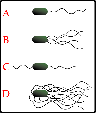
Most bacteria use their flagella to swim. These are long proteinous threads which look a little like tentacles. Some bacteria just have on of them, while others have most of their body covered in flagella, in order to provide maximum propulsion:


Flagella act like a rotory motor at the back of the bacteria, propelling it forward. Flagella based movement is usually referred to as swimming, and is the best studied of all bacterial movements. When all the flagella are moving in the same direction (usually clockwise) they shoot the bacteria forward. In order to stop, the flagella are sent spinning anticlockwise, breaking up the shape and leading to the bacteria tumbling around directionlessly, before the flagella are activated again.

However many bacteria are capible of movement without using flagella, such as the plant and insect pathogen Spiroplasma which moves due to the action of internal filaments. The contractile cytoskeleton is thought to function as a linear motor, meaning the bacteria moves along like a swimmer doing butterfly stroke, through generating a moving kink through the cell, propelling it forward.

Bacteria can also use smaller protrusions called pili, which stick out from the cell surface. There are several different kinds of pili which have different functions (type three are used for pathogenicity, as covered here) and type four pili are commonly used for movement. This is often referred to as 'twitching' as it results in jerky movement. Pili are also used to glide accross surfaces, or even to stand up and walk across them, on little pili legs!

Gliding motility does not always require pili; the bacteria Flavobacterium uses molecules called adhesins to grip to a surface and slide along it. The movement is powered by motors of Gld protein in the bacterial cell wall, as shown in the diagram below (from the reference):


Myococcus Xanthus (which I covered in further detail here) is more likely to use a motor within the cell to create a rotational movement (although movement via polysacharide secretion has also been suggested). The cytoplasmic AglZ protein is thought to act as the motor, as it remains stationary with respect to the surface as the cells glide accross it. This is shown below (image from the reference):


In addition to these active forms of movement, some bacteria also proceed more passively, without using much of their own energy. Aquatic bacteria can use internal gas vesicles in order to rise to the surface (to be closer to sunlight or nutrients). Other bacteria spread simply by growing, pushing bacteria near the surface of the colony into new terratory by all the bacterial growth around it. One of my favourite is the method used by intracellular parasites such as Listeria monocytogenes which polymerises the actin molecules of it's host behind it to travel through the human cell, leaving little superman trails of actin behind it.

There is a huge diversity in the way bacteria move, and interestingly, many of these strategies are also connected to other intracellular processes, after all the mechanism for moving yourself along a surface need not be different to the mechanism for moving molecules around directly under the cell wall. Studying these different ways helps to give an appreciation of the world bacteria live in, and how they organise themselves to survive it.

---

Jarrell KF, & McBride MJ (2008). The surprisingly diverse ways that prokaryotes move. Nature reviews. Microbiology, 6 (6), 466-76 PMID: 18461074

---

Follow me on Twitter!

Chopping bits out of the genome

ResearchBlogging.orgGenerally bacteria genomes tend to be fairly minimal in the amount you can remove from them. Unlike eukaryotes, which can have whole swathes of genome that codes for very little, bacteria, with their limited space for a chromosome, need every gene they can get. They just don't have the space for unnecessary genes.

Streptomyces bacteria, however, have bigger genomes and the luxary to invest in genes which are not strictly necessary for bacterial survival. These are called Secondary metabolite genes (as opposed to the necessary primary metabolites) and they code for genes that form an arsenal of weapons for the Streptomyces to deploy. Most Strep are soil-based, and they need the ability to produce secondary metabolites (such as antibiotics) to fight off invading bacteria, and clear terratory to expand their growth into.

What has recently been done (very ingeniously) is to remove the secondary metabolism genes from the bacterial species Streptomyces avermitilis creating essentially an 'empty' strep bacteria, that can grow and divide but not produce any of the secretory substances that strep are known for. The researchers managed to cut an entire 1.5Mb of DNA right out of the genome - helped by the fact that all the secondary metabolite genes cluster together on one side of the chromosome.

They did this using a common molbio technique, the cre-lox system. "Lox" is an area of DNA and "cre" is the protein that very specifically cuts DNA at the Lox site - it acts like a pair of scissors. They put a Lox site on either end of the DNA that codes for secondary metabolites (using a techinque called recombination in order to attach the lox into the chromosome) along with the DNA for the Cre protein under an inducible promoter. Once the bacteria had grown, they activated Cre production, which then cut the unwanted DNA out of the bacteria. This technique was amazingly successful and is shown diagramatically below (picture from the reference):


The avimitilis now contains no secondary metabolites at all, which makes a wonderful 'empty' system to use for studying how secondary metabolites are made. Genes from other bacteria, or even some of the removed genes, can be added back in, piece at a time, to see how much of the gene is necessary for metabolite production, and which regulatory pathways are the most important. Creating an empty cell also has potential implications for biotechnology, after all when trying to produce antibiotics you want the cell working as hard as possible just to produce your product, not wasting time and resources on other metabolites!

I'm really impressed at this kind of large scale synthetic-biology. It may be an area I end up going into in the near future...

---

Komatsu M, Uchiyama T, Omura S, Cane DE, & Ikeda H (2010). Genome-minimized Streptomyces host for the heterologous expression of secondary metabolism. Proceedings of the National Academy of Sciences of the United States of America, 107 (6), 2646-51 PMID: 20133795

Bacteria using bacteria

Editor's Selection IconThis post was chosen as an Editor's Selection for ResearchBlogging.orgThere are lots of things I enjoy about studying bacteria. I love their biochemistry and the secret inner workings of their metabolic pathways. I love that everything they do the manage within the confines of a single cell, and I love that you can go in there with a wrench and hit some genes until they make what you want.

But what I'm really enjoying exploring at the moment is more ecological bacteriology; how bacteria interact with their environment. How they respond to changes to stresses and, most importantly, to other bacteria. In my last post I covered how natural throat bacteria can help destroy dangerous pathogens such as MRSA Staph aureus so today I'm going to look at almost the opposite; how some bacteria can give each other a helping hand in order to infect humans.

Campylobacter jejuni is a bacteria that I feel a special affinity for because I've worked with it, back in my first ever summer project. Unfortunately it's not a very nice bacteria and can lead to bad stomach illnesses with some rare but quite threatening complications. It's found in chicken meat and cheese as it is perfectly capible of surviving happily in animals without causing them any diseases.

One of the problems with working with Campylobacter jejuni (henseforth shortened to Campy which is what we called it in the lab) is that it's very fussy about the amount of oxygen it's in. Campy is microaerophilic, which means it needs oxygen, but only small amounts, give it too much and all the cels die on you. This problem was solved in the lab by using tightly sealed containers and special packs of ... stuff ... which were put inside the containers to create the right conditions. But this does raise an important question; if the bacteria is so difficult to culture on a plate in the lab then what the hell is it doing surviving on the surface of chicken meat!

A recent study (reference below) found what you've probably guessed if you were reading this post closely, the Campy were being aided by the surrounding bacteria. The picture below shows both Campy and a bacteria called Pseudomonas putida in close interaction, with long fibrelike structures connecting them. Noone seems to be really sure what the fibre-like structures are, they may be being used for chemical communication, or they may just be keeping the bacteria in close physical contact.

The campy is the more slender and slighly spiral shaped bacteria in the centre, the others are Putida. Image from the reference.

Both bacteria were identified as being in close contact, as well as being seen together under the microscope. Further experiments were done to show that the Putida was required for Campy survival - different Campy strains were grown in both the presence and absence of the supporting Putida to see how long they could survive in completely aerobic conditions. The results are kind of hilarious, without the help of the Putida bacteria the Campylobacter just die, really quickly (image from the reference):

Figure A (top) shows the Campy with Putida grown as well, Figure (B) shows the Campy grown alone. You don't really have to be particularly good at science to interpret that one! Interestingly it was found that the interaction between different strains of both Campy and Putida was fairly specific as well, as you can see in the graph above, only three of the Campy strains have survived past 50 hours with the help of this particular Putida. Three of the Campy's still die, although they surive longer than with no help at all.

As Putida are areobic, the most likely explanation for how they are helping is that they create a microaerophilic microenvironment within their immediate surroundings. This is the kind of environment that it is thought Campy will naturally migrate to. This might be less of a helping relationship and more of a seriously exploitative one, with the Campylobacter swarming as quickly as possible towards the environment created by the Putida and then wrapping them all up in a sticky mesh to stop them moving away.

This special relationship is not applicable for all Campys, in other environments such as in humans and in chicken poo the Campy exist fine on their own, but in the highly aerobic environment of the meat surface they rely on other bacteria to survive. The implications for treatment of bacteria are intreguing (especially for antibiotic resistant strains of Campy) but it is another reminder that despite laboratory conditions bacteria do not just exist in isolation. They inhabit a whole tiny world, with challenges of it's own, surrounded by other bacteria that change their envirnment both for better and for worse.

---
Hilbert F, Scherwitzel M, Paulsen P, & Szostak MP (2010). Survival of Campylobacter jejuni under conditions of atmospheric oxygen tension with the support of Pseudomonas spp. Applied and environmental microbiology, 76 (17), 5911-7 PMID: 20639377
---
Follow me on Twitter!

Throat bacteria that destroy invaders

ResearchBlogging.orgI did a post about a week ago, talking about the relationship between the bodies natural (commensal) bacteria and the immune system. I was quite excited therefore to find a paper (reference below) which found a specific protease enzyme that is used by commensal throat bacteria to prevent harmful biofilm formation by Staphylococcus aureus, the bacteria responsible for MRSA.

The helpful bacteria in question is Staphylococcus epidermidis which lives naturally in the throat and nasal cavity of humans. When culturing these bacteria along with the Staph aureus it was found that some epidermidis cultures were capable of destroying biofilm formation, by using the protease Esp. The diagram below shows the effects of extracted Esp on colonies of Staphylococcus aureus (image from the reference):

Figures g and j show Gram stains of the colonies, the blue dye has just stained where bacteria are present. The remaining figures show scanning electron micrographs of the colonies taken at two different levels of magnification. For those interested, the scale bar for g,h,j and k is 10um and for i and l is 1um.

To double check that this protein was having an effect within the bacteria knockout mutants were made which removed the gene from Esp from the epidermidis. These bacteria were incapable of destroying Staph aureus growth. Adding a plasmid containing the Esp gene back into the bacteria restored their ability to fight off the Staph aureus which seems fairly conclusive. Furthermore this affect also works with VRSA and MRSA; Staph aureus which are resistant to antibiotics.

Below is a diagram of the effect of actual epidermidis bacteria on Staph aureus colonies (image from the reference).
These are nasal swabs taken from volunteers who had Staph aureus infections and were given the commensal epidermidis strains to try and clear them. It can be seen that the number of staph aureus is decreasing, although some bacteria are still present after five days of treatment. That might not necessarily be a bad thing as it allows the immune system to kick in with a response, and make antibodies ready for the next potential attack.

There are several exciting things that come out of this. Firstly the use of purified Esp as a defence against MRSA biofilms has the potential to be of major importance, although there may be clinical reasons why it's not such a good idea to spray proteases all over the inside of someones nose! From a less medically-useful perspective it's a wonderful example of bacterial-colony interaction. The kind of struggle for survival that happens inside your nose is occurring for bacteria everywhere; in soil, in the water, in the air, and even in humans.

From the Staph epidermidis point of view your nasal cavity is just a great place to live (warm, safe, lots of nutrients) and it's not going to give up that kind of living environment without a fight!

---

Iwase, T., Uehara, Y., Shinji, H., Tajima, A., Seo, H., Takada, K., Agata, T., & Mizunoe, Y. (2010). Staphylococcus epidermidis Esp inhibits Staphylococcus aureus biofilm formation and nasal colonization Nature, 465 (7296), 346-349 DOI: 10.1038/nature09074

---
Follow me on Twitter!

The Gibson Assembly Song

There are no words to describe how awesome this is. You just have to watch it:


This is The Gibson Assembly song, written by the Cambridge iGEM team 2010. If you can't see it, want it in a higher resolution or just want the link to share, find it here.

The technique they are describing is the "Gibson Assembly" which is a fairly quick and painless way of joining two bits of DNA. In more sciency terms, it works by using PCR to make genes with large overlaps (40bp) at the end. You add a Master Mix to the fragments, incubate for one hour, then just transform into whatever cell you're using. For more details of how it works go here, for the recipe of the Master Mix and detailed protocol go here, and for a program to help you design Gibson Primers, go here.

The video was just made for fun, and took less than a week to put together (not counting the time to write the words!). Filming was done mostly over one day, using one camera and a slightly broken tripod, just using spaces in the lab and the gel room. The green-screen sections were done by throwing a green table cloth over some poster display boards. The music was recorded seperately, and I think each instrument was recorded seperately as well, to get the sound balance right. It was all carried out by about nine undergraduates (and one lab rat!) and massively confused most of the supervisors.

I'm already starting to see some replies and responses to this, the most popular ones seem to be variations on the "why aren't you doing any real work!?!" and "How do you have the time with all your Science!" And given that there's been a lot of kurfluffle in the blogosphere lately about science, time spent doing science and the connection of both with Passion for science I suppose I should address this...

The undergrads who made this video had been working all summer, pretty much starting two weeks after term finished. They worked weekends. They worked evenings. At the time of doing the video, they'd worked several weeks over their stipend and had long ago stopped being paid.

Now their term has started, and they are still coming in to work, in between lectures, in between their own projects. They also have to prepare a presentation and a poster over the next month, all in between their term work. And terms here are manic; eight weeks of non-stop craziness that it's hard to fit anything into.

It was just a quick few days of Fun, in between the Science. :D

Friend or foe? How the immune system copes with the gut microbiotica

ResearchBlogging.orgThe job of the human immune system is to destroy pathogens. Using a combination of quick, immediate responses (the innate immune system) and long-term memory (the adaptive immune system) in humans the cells of the immune system are perfectly primed to seek out any cells that are Other (i.e not Self) and kill them.

Which leads to a slight problem, because rather a lot of the cells within your body are 'Other' cells, and their existence is vital to your health. Within your stomach, and your respiratory tract, live a number of commensal bacteria, friendly and harmless bugs that can survive quite happily inside you and help to fight against incoming pathogenic bacteria. Stripping away the bacteria in the gut (i.e by going on a course of very strong antibiotics) leads to all kinds of problems including digestive problems and, once the antibiotics have finished, increased risk of disease-causing bacteria invading the now bacteria-free stomach.

In fact several notable yoghurt making companies are making a lot of money by selling you drinks with bacteria in them. They reassure you that the bacteria aren't dangerous, which is all well and good, but they never quite explain why the ingestion of many bacteria doesn't cause your immune system to have a panic attack.

A new review in Nature looks at the interactions between the gut microbiome and the immune system. The 'gut microbiome' is the collection of bacteria that start colonising the inside of your intestines soon after birth, both from your mother, and from the general environment. It's helpful here to remember that technically your intestinal tract isn't actually inside your body. There's an open tube right the way from your mouth to your arse (for want of a better word...) so the body has a tendency to treat bacteria living there in similar ways to the bacteria living on your skin, by using barriers to keep them out.

However there still is a trade off. The cells that make up the intestinal walls still need to be able to respond to bacteria, and the commensal bacteria still need to be contained. A non-regulated population of bacteria will simply keep growing until all available space is filled (and all nutrients eaten), and this does not happen within the gut.

Starting with the innate immune system which works by recognising molecules found in all pathogens (called PAMPs) these are recognised by human cells using receptors called TLRs (Toll-like receptors - long story) and lead to a signalling cascade that result in a huge number of cytokines and other inflammatory agents being released to kill the bacteria. In the gut this wouldn't just lead to the massive slaughter of the microbiome, but also to a huge amount of damage to the surrounding human cells. Enough exposure to microbial elements such as lipopolysacharrides can downregulate this response; the lipopolysacharrides (which are in the bacteria cell wall) down-regulate one of the key components of the signalling system, a molecule called IRAK1. This prevents the cell from mounting a response to the bacteria. For those that want scientific details, check out the diagram below (image from the reference):


The adaptive immune system is more complex. In normal situations it works by taking a small sample of the bacteria back to the lymph nodes and preparing a specific immune response against it. Special immune cells (B cells) are then made which will kill the specific bacteria, with the help of T cells, which also act as a memory of the threat and the correct response. The B cells are then sent to the point of infection and secrete antibodies which clump the bacteria into groups and recruit other factors to kill them.

This still mostly happens in the case of the gut microbiome, the B cells release the antibody IgA which diffuses out into the intestinal tract and traps the bacteria in the mucus layer. However the bacteria are able to strike back, not by targeting the B cells, but the T cells. There are many different forms of T cells, and by secreting certain chemicals the bacteria can encourage the formation of T-regulatory cells which encourage tolerance towards both commensal bacteria and molecules in food.

So it seems to be not so much a relationship of mututal tolerence and understanding, but more like a sort of uneasy standoff. Bacteria are still being killed to stop them spreading, but are holding off the immune systeme enough to maintain a steady population. In return, the immune system is still there and active, but not active enough to cause any serious damage to either the microbiotica or the surrounding human cells.

---

Cerf-Bensussan N, & Gaboriau-Routhiau V (2010). The immune system and the gut microbiota: friends or foes? Nature reviews. Immunology, 10 (10), 735-44 PMID: 20865020

---

Follow me on Twitter!

Bacterial Fossils

ResearchBlogging.orgStudying ancient bacteria can be a little difficult. Despite having wonderfully complex internal biochemistry and fascinating ecological interactions they are essentially, when you get down to it, a little soggy bag. Little soggy bags do not preserve all that well; when bacteria die they just break apart, and are often eaten by other scavenging bacteria which see them as just free nutrients.

However some bacteria do fossilise, leaving behind perminant records of their existance. One of the ones that does it the best is cyanobacteria, which can form fossils in two ways. Firstly by forming little calcified shells around themselves as a product of increasing the dissolved carbon dioxide levels inside the cell:

Cyanobacteria fossils - from Berkeley university page

Secondly cyanobacteria can group together with algae to form large layered mat-like structures called stromatolites. Slicing these very thinly reveals tiny cyanobacteria fossils, caught between the layers. There are stromatolytes in Australia that have been alive and growing (very slowly) for millions of years, despite looking faintly uninspiring.

Maybe not quite as impressive as dinosaurs...

While no other bacteria form fossils as nicely preserved as the cyanobacteria, they are capable of leaving behind visible remnants of their existence. The polypeptides of the bacterial cell wall (along with cytoplasm and some secreted lipids) can, under certain conditions, act as nucleation sites for minerals. This eventually leads to the organic cell being replaced by a little mineral cast of the bacteria. There is the possibility for quite a few artifacts with this (artifact being the scientific word for "result caused by the preservation process rather than the bacteria") the most common one being the creation of an artificial nucleus structure. As the bacteria degrades the cytoplasm tends to clump, and the crystallisation of minerals around a cytoplasm clot can in some cases create a structure that looks similar to the structure formed by a nucleus.

Endolithic bacteria that live in rocks can leave behind tiny canals in the rock surface that they bore into. These are a lot harder to find and interpretation is usually helped by the discovery of nearby alive endolithic bacteria. Bacteria have also been found trapped and mumified inside tree resin, a la the Jurassic park mosquito.

Once you start getting larger organisms the bacteria have a brand new niche to exploit. Fossilised bones sometimes show the results of a bacterial infection; while the bacteria themselves are not being preserved their presence is still seen in the fossil record.

Unfortunately although these glimpses are really interesting their also kind of frustrating from a biochemical point of view. They provide clues as to the lifestyles and processes within the bacteria but they are such tiny clues. Past biochemical processes are more often found inside the actual bacteria, by looking at clues in the genome and the genomes of related bacteria, than they are in the remains the bacteria leave behind.

---

Perri, E., & Tucker, M. (2007). Bacterial fossils and microbial dolomite in Triassic stromatolites Geology, 35 (3) DOI: 10.1130/G23354A.1

Westall, F. (2001). Early Archean fossil bacteria and biofilms in hydrothermally-influenced sediments from the Barberton greenstone belt, South Africa Precambrian Research, 106 (1-2), 93-116 DOI: 10.1016/S0301-9268(00)00127-3

Guest Post - Survival of the fittest?

ResearchBlogging.orgI'm very excited about this post, which is a guest post from my sister! She's an undergrad doing biochemistry at Bristol University, and she's currently taking a year working in a research laboratory as part of her degree. She's working with Plasmodium at the moment (which is the little protist that causes malaria) but has sent me a bacteria-related post because she knows me, and she knows my blog and who doesn't love bacteria?

Post - survival of the fittest?

Bacteria have always been very adaptable when it comes to surviving evolutionary stressful situations, such as exposure to antibiotics. Usually some form of mutation will arise leading to the creation of resistant strains of bacteria. These will be selected for via ‘natural selection’ processes and go on to replicate to produce a whole population of resistant bacteria that are able to survive.

However new research looked at a colony of wild type E.coli bacteria in a bioreactor under exposure to increasing levels of the antibiotic norfloxacin and found that no more than 60% of growth was inhibited to maintain a sizable population. The resistance levels of the population as a whole, and of 12 random individuals, was checked every day and it was found that they did not correspond to one another.

The majority of the individual isolates were less resistant than the population as a whole but there was one mutant found that was highly resistant. By isolating the supernatant from the high resistance individual, and conducting gel electrophoresis to separate out the intracellular components, a protein was found that was produced in very high numbers.

This was the enzyme tryptophanase which has the main job of breaking down tryptophan to ammonia, pyruvate and indole. Experiments were done to show that the third molecule, Indole, provided an obvious survival benefit under antibiotic conditions. It upregulated multi-drug efflux pumps which helped in the physical export of the drug and it also had a role in activating various oxidative stress protective mechanisms. The mass production of Indole by the highly resistant mutant allows more vunerable cells in the surrounding area to survive.


a) no antibiotic stress, bacteria naturally produce indole.
b) dead and dying bacteria fail to produce indole
c) mutant appears and supplies indole at a fitness cost to itself.

The resistant bacteria is therefore not selfishly replicating to outgrow the rest of the population but, in helping others to survive, is enduring a fitness cost of its own by mass producing Indole.

This experiment was also carried out using various different antibiotics and the same bacterial altruism was found to exist. The survival of the weaker bacteria does have some advantages as it allows further exploration of mutations that could be even more beneficial to the population. Also, it keeps the opportunity for the bacteria to return to their original state if the stress is temporary, rather than keeping up the energetically wasteful production of antibiotic resistance genes.

So, bacteria working as a team to ensure not just temporary survival but long term advantages for the whole population. Not just survival of the fittest.

---

Lee HH, Molla MN, Cantor CR, & Collins JJ (2010). Bacterial charity work leads to population-wide resistance. Nature, 467 (7311), 82-5 PMID: 20811456

I for one welcome our glowing overlords...

I've already shown you pictures of the glowing bacterial lightbulbs, but in terms of glowing products the cambridge iGEM team is going from strength to strength. To start with the most amazing, they've made an oxygen activated bacterial bubble-lamp:


(It can be found here if the above does not work)

Maybe it's not the most efficient thing to read by, but it's an impressive level of brightness and it turns off relatively (biologically speaking!) fast. It's simply a large measuring cylinder with a load of bacterial broth inside it, and a tube to blow air through. Even shaking lets enough oxygen through to start turning on the light.

Another thing they've been doing is playing around with images on 24-well plates. For those who haven't used them, 24-well plates are usually run on a plate-reader which reads samples from every well, used mostly (in our lab) for overnight assays or (as I should be doing tomorrow) as a glorified spectrometer, measuring a range of absorbances over different wavelengths.

The iGEM team are using them to make nerdy pictures. This is my favourite:

The thing I like about the 24-well-plate pictures is that their different. Painting on plates is awesome buts it's been done before a couple of times, and the glowing pixel-pictures just look new and fresh and exciting.

There's probably going to be a bit of speculation as regards this of the "but how useful is it" type. And rest assured the iGEM team are thinking of that but at the moment I'm happy to just enjoy the fact that we have glowing pixelated space invaders sitting on the bench.

Glowing pixelated space invaders!

This is why I went into science :p