Field of Science

Showing posts with label papers. Show all posts

Vaccines and viral evolution

ResearchBlogging.orgThis is the first time in ten years that I haven't had an exam around summer-time. It feels odd, everyone around me is either exam-stressed or post-exam-relaxed, it's turning to summer and there's a definite final term feeling but this time I'm not really part of it. It's been an interesting year this year, since January I've not been involved in any part of research science, other than writing about it.

However luckily I'm still surrounded by lectures, seminars, talks and various other interesting stuff which no one seems to mind me occasionally turning up too. Seeing as I haven't written much about virus's lately I headed over to a talk the other day about Marek's disease, which is caused by a Herpes Virus and has a rather devastating affect on chickens.
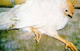
It makes them stand like this :(

It started back in the sixties, when rather a lot of chickens suddenly started dropping dead, sometimes up to 50% of all the stock in a large barn. Bear in mind these weren't the happy pecking-around-shrubs-of-grass chickens that feature on the front of free-range eggs, but rather a lot of chickens quite closely packed inside a big barn. The cause was found to be MDV - Marek's disease virus. After a lot of work a vaccination was found and given to all the chickens. Over $2 billion was saved by this, and the chickens were able to survive, right up until they got slaughtered for food.

But then, around the 1980s the disease suddenly reared it's head again, this time in a far more virulent form imaginatively labelled vvMDV (which stands for very virulent MDV). More research, another vaccine, and the deaths stopped.

Until just before 2000 when the virus evolved again into an even more virulent strain called vv+MDC, which means exactly what you think it does. Another vaccine was made (called Rispens) but at this point it was becoming fairly clear that this virus was behaving oddly. Three times it had changed, becoming more and more deadly each time:

Image from the presentation slides showing the development of new strains with an increase in virulence.

This is not normal behaviour. Virus's rely on their hosts, they can't replicate, survive or do anything without a host cell, which means they have a vested interest in keeping the host alive. If anything, viral strains should evolve to become less virulent; a virus that kills the host will be a virus without a host and is therefore less likely to survive and propagate than a virus with a host.

It turns out in this case that the evolution of increase virulence is down exclusively to the way the vaccine interacts with the virus. The virus works by being inhaled into the chickens lungs, getting into the cells of the immune system (B and T cells) and causing a latent infection of the lymphocytes (T cells). Virus cells can also work their way to the epithelial cell of the feather follicles and will shed from under the feathers, thus keeping the virus in circulation.

In an ideal situation a vaccine would produce what is known as "sterilising immunity", where use of the vaccine kills all viruses dead. This is how almost all human vaccines work. With the Marek's disease vaccine however, the virus was not killed completely, but could still replicate and shed from the feathers. This means that the virus was still within the system, able to change and evolve. The vaccine however, does make the virus less likely to spread, which means that a more virulent form that the vaccine does not protect against is able to outcompete the less virulent strain. Because the chickens are all in very close proximity, and because there are a lot of them, the more virulent strain can spread much faster throughout the population. With normal chickens, in small isolated populations this would not happen as a virus that virulent would run out of host and die out.

This creates a paradox - vaccines are needed to stop the chickens getting the virus, but at the same time use of the vaccine is creating an evolutionary environment in which a more virulent virus can grow. There are some responses to avoid this though. Firstly, to develop a virus that produces sterilising immunity, i.e that kills all thee virus dead. Secondly, to allow the chickens more room and freedom to stop the virulent strains spreading so quickly. And while this second solution sounds like every animal-rights campaigner's dream, remember that it only really works if very few people in the world eat chicken. In reality there are lots of people, and a limited space for chickens - eating chickens which have lead a happy healthy life is a privilege for a few, not the reality for the majority.

---
Witter RL (2001). Protective efficacy of Marek's disease vaccines. Current topics in microbiology and immunology, 255, 57-90 PMID: 11217428

Witter RL (1997). Increased virulence of Marek's disease virus field isolates. Avian diseases, 41 (1), 149-63 PMID: 9087332
---

Follow me on Twitter!

Social Evolution in Bacteria - SGM series

This is the fourth post in my latest SGM series.

ResearchBlogging.org
The social behaviour of bacteria is something that I get very excited about. From the wolf-pack hunting strategies of Myxococcus xanthus to the terminal differentiation of cyanobacteria, it's something that I never get tired of writing about. As well as providing interesting quirks of bacterial behaviour, living within a colony also gives new scope for exploring the evolution of bacteria; not just as single entities but as a fully functioning social group.

One of the differences of living within a social colony as opposed to alone means that altruistic-type behaviour has to be adopted. Bacteria living within a biofilm need to excrete the sticky goo that holds the biofilm together, which is problematic because synthesising and secreting goo takes up a lot of energy. So within this colony, there will be 'cheaters' - those bacteria that live in the surrounding goo produced by others, while making none themselves.

A bacterial biofilm, showing individual bacteria in green. Image taken from the FEI website, shown there courtesy of Paul Gunning, Smith & Nephew

As with all colonies, cheating might benefit the individual but has no benefit for the colony as a whole. Too many cheaters and there won't be any biofilm. And recently an even more subtle form of cheating has been shown within the biofilms of the bacteria Pseudomonas aeruginosa, with bacteria that don't just refuse to make vital sticky chemicals, but also abstain from the entire process of forming a biofilm.

Bacteria use a complex communication system called quorum sensing in order to determine how many other bacteria they are surrounded by. Once enough bacteria are present, all signalling their existence, the biofilm will start to form. However some bacteria isolated from the biofilm were shown not to be taking part in any quorum sensing at all. Quorum sensing appears to be quite a burden for a growing cell - cells with the quorum sensing genes knocked out tend to grow a lot faster that the socially conscious cells that allow biofilms to form.

The paper that goes through this (reference one) highlights it as a form of social cheating, with bacteria avoiding quorum sensing to benefit themselves while mooching off the quorum sensing behaviour of others. I'm not entirely certain that this is the case though. It may just be an good example of job allocation within the bacterial society. Clearly not all bacteria are required to be continually quorum sensing, so why should they all have to? Would it not be more sensible to have some exempt from that task, so that they can concentrate on growing, dividing, and spreading the colony? This may be more a case of tax-breaks than of benefit-cheats.

Social evolution doesn't just take place within species, but also between them, and like every other organism bacteria are in a constant state of coevolution with both their 'prey' and their predators. Most predator-prey interactions take long periods of time to study, but the beauty of bacteria is that you can go through several generations in the course of one week's growth. Studies of the bacteria Pseudomonas fluorescens and its bacteriophage parasite showed that both the bacteria and the bacteriophage evolved far quicker when interacting together than they did when competing against a non-changing opponent.


Bacteriophage surrounding a bacteria. Image from wikimedia commons

'Evolve' here means that the bacteria and the bacteriophage showed a greater change in their genetic makeup, and a greater genetic divergence from bacteria not pitted against the phages. Unsurprisingly, the genes that changed the most were those involved in host-phage interaction. This study (reference 2) is also a great example of the usefulness of whole genome sequencing. Whole populations of bacteria and phage were allowed to evolve both together and separately and then just sent away for sequencing with the results analysed at the end.

You really can't be an anti-evolutionist while studying bacteria. They just do it so damn quickly and often you can see it happening.

---
Sandoz, K., Mitzimberg, S., & Schuster, M. (2007). From the Cover: Social cheating in Pseudomonas aeruginosa quorum sensing Proceedings of the National Academy of Sciences, 104 (40), 15876-15881 DOI: 10.1073/pnas.0705653104

Paterson S, Vogwill T, Buckling A, Benmayor R, Spiers AJ, Thomson NR, Quail M, Smith F, Walker D, Libberton B, Fenton A, Hall N, & Brockhurst MA (2010). Antagonistic coevolution accelerates molecular evolution. Nature, 464 (7286), 275-8 PMID: 20182425
---

Follow me on Twitter!

Signals for Infection

ResearchBlogging.orgNeisseria meningitidis is a bacteria which lives in the throats of around 30% of the human population. In most cases it causes no problems at all and just exists as a normal part of the throat microbial flora. In some patients however it can start to colonise the bloodstream and brain, leading to cases of septicemia and meningitis which are highly dangerous and can be fatal.

The invasion starts with individual bacteria, which adhere to the epithelial cells that cover the inside of the throat. They then start to divide and proliferate to form large aggregated colonies. Within these colonies they are connected to each other, and to the epithelial cells, by protrusions from the bacterial cell surface called pili which are shown below for a wild-type (i.e un-genetically modified) Neisseria meningitidis:
Image taken from the reference below. The arrow points to one of the pili, and the insert shows a close-up of it.

These pili are often modified by the attachment of small molecules to the pili proteins, including the molecule phosphoglycerol (shown on the right for those interested in structure). To test the effects of the addition of phosphoglycerol, the researchers found which gene caused the addition of this molecule onto the pili (the pptB gene), and removed it from the cell. Without the pptB gene there was still the same number of pili around the cell, but they were not clumping together as much. Instead of the thick fibres seen in the wild type above (caused by large bundles of pili) only little stringy fibres were seen. These thin spindly fibres show that without the addition of phosphoglycerol, the pili cannot clump together.

This is important medically as Type IV pili bundle formation and N. meningitidis aggregation for infection are linked. Interestingly it was not the aggregation that was affected by removing the phosphoglyerol but the ability of individual bacteria to leave the aggregate to infect other parts of the body. In wild-type bacteria, the pptB gene is strongly activated only after several rounds of division within the aggregate, so it looks like the addition of phosphoglycerol acts as a switch, communicating to the bacteria that enough of them have aggregated and it is now time to leave. If the pptB is activated due to large numbers of bacteria it could act as a communication of the population density - signalling to the individual bacteria that the current location is far too crowded, and it has better chances of survival if it leaves.

---
Chamot-Rooke J, Mikaty G, Malosse C, Soyer M, Dumont A, Gault J, Imhaus AF, Martin P, Trellet M, Clary G, Chafey P, Camoin L, Nilges M, Nassif X, & Duménil G (2011). Posttranslational modification of pili upon cell contact triggers N. meningitidis dissemination. Science (New York, N.Y.), 331 (6018), 778-82 PMID: 21311024
---
Follow me on Twitter!

Multicellular signalling

ResearchBlogging.orgI like studying bacteria. I find them fascinating, wonderful little creatures, able to do as much (and often more!) with a single cell as other organisms need whole multicellular bodies to achieve. I like exploring the places bacteria live, the things they can do, the ways they manage to exploit practically every niche on earth, and of course most importantly how I can exploit them.

But not everyone loves bacteria, and at heart I am a biochemist which means, among other things, that I get to teach younger biochemists. This means I do occasionally find myself venturing uncertainly into the world of the multicellular and while doing so recently I found an interesting paper on cell signalling (reference below) which I thought I would share.

All cells need to be able to communicate, but while bacteria know that everyone they communicate with is a competitor, multicellular organisms have cells that need to be able to cooperate in a strange and slightly twisted form of cellular-communism. Each cell needs to know when it can divide (usualy never), when to grow, when to release chemicals and, ultimatly,when to sacrifice itself for the Greater Good.

Cellular communication is mostly a chemical affair, with small molecules called ligands being sent from one cell to another and recognised by receptors on the cell surface. These receptors can take many forms, but one of the more common ones is the form of a seven-transmembrane spanning receptor, so called because it goes through the membrane seven times:

Picture (c)me and my dodgy art skills. The protein is in blue, the membrane in pink, and the ligand bound on the outer cell surface is the red blob.

Binding of a ligand causes a conformational change in the whole structure, most importantly in that long intracellular tail shown above. This can then activate other molecules inside the cell, with the end result that a specific gene is turned on or off. In the classical model of this process the intracellular tail interacted with a little molecule called the G protein which carried the message through to the genome. Another protein that featured in this model was B-arrestin, which was thought to desensitise the receptor and the G-protein by re-setting it back to its original state, i.e switching the thing off. This model is shown below:

Picture (c) me. This is a simplified diagram, in 'reality' there are a lot more different proteins involved, but these are the main ones, and the important ones for this paper.

New evidence is coming to light which modifies this model. Firstly, it's been found that the B-arrestin does more than just switch off the G-protein, it is also capible of sending its own signals, through a cascade of different proteins. Both the G protein and the B-arrestin can be used to pass on the message sent by the ligand. Secondly, it's been found that these two proteins are not activated equally, a bias can be displayed, sending the signal through one of these two intermediate proteins; either the G protein, or the B-agonist or a mixture of the two. This bias can be either due to the properties of the receptor, or those of the ligand binding to it. Experimentally you can generate a bias by altering either the receptor or the ligand to prefer binding to the B-agonist, and you can plot these on mathematical-looking graphs.

You can tell this is a biology graph because there are no actual numbers, just vague concepts :p (c) me.

The actual physiological effects of this are only starting to be explored, as it introduces an extra level of complexity to intracellular control. The use of several different ligands, all with varying degrees of bias at the same receptor, could produce more subtle cellular output responses. Within a multicellular organism, the better your intracellular communication is, the more likely your organism is to grow happily and survive.

---
Rajagopal S, Rajagopal K, & Lefkowitz RJ (2010). Teaching old receptors new tricks: biasing seven-transmembrane receptors. Nature reviews. Drug discovery, 9 (5), 373-86 PMID: 20431569
---
Follow me on Twitter!

Guest post - Microbes and Madness

This guest post comes from my fiancé who is a Psychiatrist. I've been very excited about this post for a while, because unlike me, he is a published author who has written a book on Consciousness and the philosophy of mind.

Microbes and Madness

At first glance, it would be reasonable to assume that my profession and that of the author of this fabulous blog are poles apart. However, everything in nature has a connection, and so it is not surprising to discover a fascinating area where psychiatry and bacteriology overlap.

A broad range of pathogens are known to cause psychiatric sequelae, including worms (neurocysticercosis), protozoa (cerebral malaria, toxoplasmosis), viruses (HIV, herpes simplex encephalitis, rabies), prions (Creutzfeldt-Jakob disease, kuru), and, of course, bacteria (neurosyphilis, Lyme disease, post-streptococcal syndromes). However, in the spirit of this blog, this post will be focusing on bacteria.

There are essentially four mechanisms through which bacteria cause psychiatric symptoms in humans:

I. Bacteria can infect the central nervous system and cause direct damage to brain cells.
II. Bacteria can trigger a powerful systemic inflammatory response that results in a disruption in brain function.
III. Bacteria can trigger an adaptive immune response which produces antibodies that cross-react with host central nervous system proteins.
IV. Bacteria can be the objects of a phobia.

Syphilis and Lyme disease are examples of infections which involve the first mechanism. Syphilis is caused by spirochaetes of the species Treponema pallidum, and is sexually-transmitted. Lyme disease is caused by spirochaetes of the genus Borrelia, and is vector-borne, with ticks from the genus Ixodes being the commonest vector. Both diseases are associated with widespread dissemination of infection of multiple organ systems, and are notorious for their protean manifestations.

The range of possible psychiatric presentations is vast. Syphilis, in particular, can mimic any psychiatric syndrome, and was a common diagnosis in psychiatric inpatients a century ago. The possible range of presentations include delirium, dementia, psychosis, mania, and personality changes. Lesions of the frontal lobes are associated with personality changes and disinhibited behaviour, whereas those of the temporal and parietal lobes are associated with cognitive decline. Lyme disease can also mimic several different psychiatric syndromes, but typically affects the limbic system, causing disorders of emotional regulation, including panic attacks, phobias, depression, and obsessive-compulsive behaviour.

The second mechanism listed refers to sepsis-associated delirium. No human organ system is a closed system, including the central nervous system. Bacterial infections with a focus outside the outside the brain are capable of causing a systemic reaction, which affects the brain. The result is an acute confusional state, or delirium.

Common causes are pneumonias and urinary tract infections, although infections of other organ systems are also frequently implicated. Delirium presents as a transient global disorder of cognition. Typically, there is clouding of awareness, disorientation, impaired attention, fluctuating alertness with agitation or drowsiness, hallucinations, illusion, and vague delusions. The state is thought to be caused by a global disruption of brain function, which may result from the effects of a systemic inflammatory response to infection. These effects may include systemic vasodilation causing cerebral hypoperfusion, increased permeability of capillaries allowing toxins to cross the blood-brain barrier, the action of inflammatory cytokines on the brain, and increased body temperature resulting in an increase in neuronal oxygen demand.

The third mechanism is seen following infections with group A beta-haemolytic Streptococcus pyogenes, such as scarlet fever and tonsillitis. In response to infection, the adaptive immune system produces antibodies against antigens on the invading pathogen. However, some streptococcal antigens are similar in some way to antigens on host tissues, and so the antibodies produced mistakenly recognise and attack the host tissues. Examples of post-streptococcal autoimmune diseases include rheumatic fever, glomerulnephritis, and Sydenham’s chorea.

A psychiatric syndrome caused by this mechanism is PANDAS, which stands for paediatric autoimmune neuropsychiatric disorder associated with streptococcus. This typically presents as a dramatic onset of obsessive-compulsive disorder, tic disorders, or Gilles de la Tourette syndrome following an infection with group A beta-haemolytic Streptococcus pyogenes in childhood. It is thought to be a result of autoimmune damage to the basal ganglia, which is the part of the brain involved with the initiation and regulation of motor commands. Interestingly, it has also been suggested that encephalitis lethargica, a mysterious syndrome which caused an epidemic during World War I, may also be caused by a post-streptococcal autoimmune reaction.

The fourth and final mechanism listed refers to mysophobia, or the pathological fear of germs. Behavioural symptoms include repeated washing of hands, excessive cleanliness, and avoidance of social contact. Anxiety and panic attacks also occur. Although the behavioural manifestations are similar, mysophobia is not to be confused with obsessive-compulsive disorder. The former is a phobic disorder, in which the fear of germs underlies the behaviour, and the function of the behaviour is avoidance of the phobic object. In the latter, the behaviour is compulsively carried out in response to the obsession that the behaviour must be carried out.

I hope to have provided an comprehensive overview of some of the interesting ways microbes can cause mental and behavioural disturbances in humans. The function of this ability is open to speculation. The film 28 Days Later tells the story of an artificial ‘Rage’ virus. When a human is infected, he or she becomes uncontrollably aggressive, attacking other humans and infecting them with viruses in the process. Thus, the viruses’ effect on human behaviour is clearly advantageous to their spread and propagation. However, outside of fiction, the advantages of pathogens’ effects on human behaviour is less obvious. Even with rabies, on which the symptoms of the ‘Rage’ were based, there has been no documented human-to-human transmission through bites. In fact, the only documented cases of human-to-human transmission of rabies were of transplant recipients receiving corneas from infected donors! It is therefore not known what evolutionary advantage, if any, the psychiatric sequelae of infection convey to the pathogens. It is possible that they are epiphenomenal.

---
Pfister D, Siegemund M, Dell-Kuster S, Smielewski P, Rüegg S, Strebel SP, Marsch SC, Pargger H, & Steiner LA (2008). Cerebral perfusion in sepsis-associated delirium. Critical care (London, England), 12 (3) PMID: 18457586

Neurosyphilis: Considerations For A Psychiatrist Mark A. Ritchie, Joseph A. Perdigao, Mark A. Ritchie

The Neuropsychiatric Assessment of Lyme Disease

A. Mazzola, G. Mazzola (2006). OCD And Beta Haemolytic Streptococcus: A Nasty Association. Priory publishing link

Oil-eating bacteria

ResearchBlogging.orgOil is formed from hydrocarbons: organic compounds which consist soley of the elements hydrogen and carbon. There are many, many different types of hydrocarbons, all of varying lengths and shapes, and pretty much all of them can in some way be consumed as an energy source by bacteria.


Hydrocarbons, in both ring and chain form, taken from a mix of sources

The general rule is that the shorter and fewer rings present, the more toxic the compound to bacteria (ethanol, for example, is deadly) however there are very few hydrocarbons that some bacteria somewhere won't consume. Like all living organisms, bacteria need a carbon source for energy, and when there are none of the coventional ones around (such as glucose and lactose) many of them will start consuming the ones in oil - crude oil contains a lot of carbon, which is why we burn it for energy in the first place.

Oil-eating bacteria have been found in a huge number of locations as well - seawater, freshwater, groundwater, water containing sludge, and land environments such as silt, soil and sand. They are found in arid deserts o the Middle East (which to be fair is a good place to hang out if you consume oil) and even in ice cores from Antarctica. And rather than being found widespread within seperate species, the capacity for oil consupmtion is often found in many different species within a particular location - in the bacterial world you are more likely to share habits with your nearest neighbours than with your closest relatives.

Using these bacteria for our own purposes, however, is proving slightly more problematic. You can purchse freeze-dried hydrocarbon-degrading bacteria and sprinkle them on your oil slick like minature Captain Planets, but their abilities are currently not nearly as effective as chemical detergents, or physical removal of the oil. This is mostly because these freeze-dried bacteria are specialised lab strains that (much like several of the academics that constructed them...) are often highly unsuited to the real world outside of the safe confines of the lab. Sometimes as well the pollutant is unaccessible to the bacteria, which may be floating on the top of the water while the oil is in droplets underneath the surface.

Looking into the potential of oil-eating bacteria is encouraging though, especially after incidents like the BP oil spill earlier this year. For the oil eating bactera that were hanging around, that must have been a feast day! And even though that event has now been largely forgotten by the media, there will still be coils and dropplets of oil floating around from it, and those will be cleaned up almost exclusively by bacteria.

---

Lena Ciric (2010). A natural solution: how bacterial communities can help clean up oil spills Microbiology Today, 229-231

---
Follow me on Twitter!

Storing DNA

ResearchBlogging.orgDNA is one of the most important components of the cell. In eukaryote cells (i.e the cells of humans and plants) it is stored inside a nucleus that keeps it safe and away from dangerous things like free radicals produced by the metabolic reactions of the cell. In bacterial cells the DNA isn't nearly as well protected, but the main bulk of the bacterial chromosome (excluding the little floating plasmids) is all kept together in a bundle usually referred to as a nucleoid.

However the DNA in cells is rather long which means that in order to package it into a small space it needs to be coiled up. Eukaryotic cells do this by using proteins called Histones, which coil the DNA around them, and can also signal which genes the cell should be turning into proteins.

Image from wikimedia

Because eukaryotes cells have so much DNA they need to keep it all tightly coiled up, with little molecular tags on the histones to remind them which bits are needed reguarly, and which bits can be mostly ignored. Prokaryotes on the the other hand don't need quite as much control, most of their DNA is going to be used most of the time, but it still needs to be packaged up to fit inside the cell.

The cells do this using by using the unbelieveably eukaryot-ist named "histone-like proteins" such as H-NS which is found in E. coli and related bacteria. Although their precise mechanisms are not quite clear, they have been shown to play a clear role in the coiling of the DNA and maintaining nuceolide stability. They also (like their eukaryote counterpart) help to control gene expression as well as chromatin structure.

In most cases, the H-NS functions as a transcription repressor merely through its physical presence wrapped around DNA - it's harder to transcribe DNA (the first step for protein production) when it's all wrapped up tightly in balls. Some genes though, seem to be activated when associated with H-NS, as it can affect the stability of the mRNA transcript. These are usually genes associated with stress responses; in fact many of the genes that are affected by H-NS are linked to the stress response or changes in the environmental conditions, such as high or low temperature, high osmolarity, changes in pH or oxygen concentration.

Quite how the H-NS controls these parts of the DNA is not entirely clear. Unlike histones, which are positively charged to stabilise the negative charge on the DNA, H-NS proteins are neutral. Although the domain of the molecule that binds to the DNA has been identified, it is not yet certain which parts of these are vital, or how they interact with the DNA molecule. One thing that is clear however is that H-NS are capible of forming dimers, and probably carry out much of their task as a dimer of two joined molecules.

Domains of an H-NS molecule, from the reference. The oligomerization domain binds to a fellow H-NS while the DNA binding domain attaches to DNA.

Whatever the binding mechanism is, it is unrelated to the actual sequence of the DNA, as H-NS can bind to many different regions of the gene, regardless of sequence. Until recently the H-NS molecules were thought to have no post-translational modifications (unlike histones which are often decorated in molecular markers to indicate which kind of gene they are on) but some have recently been found to be marked with poly-3-hydroxybutyrate, a small lipid molecule. The reason for this is unclear, but it does raise some exciting implications for H-NS control of gene transcription.

When it comes to controling DNA expression, it's clear that in both eukaryotes and prokaryotes, the scaffold proteins that hold the DNA coiled up act as more than just a scaffold. Instead they are involved in a substantial amount of the control of DNA expression, often working closely with other control proteins to ensure the correct genes are turned into proteins.

---

Schröder O, & Wagner R (2002). The bacterial regulatory protein H-NS--a versatile modulator of nucleic acid structures. Biological chemistry, 383 (6), 945-60 PMID: 12222684

---
Follow me on Twitter!

Bacteria that tear themselves apart

This post was chosen as an Editor's Selection for ResearchBlogging.orgBacterial cell division is one of those fairly well studied areas, where time and much study has come forth with a nice standard model. One of the main proteins involved is FtsZ, which seperates one bacteria into two by forming a ring of protein around the middle of the bacteria and tightening it shut as shown below (figure from Nature paper):

Until quite recently it was thought that this was pretty much the only way to get bacteria to divide, until 1999, when the sequences of two Chlamydia species turned out not to contain genes for FtsZ. As Chlamydia are intracellular parasites (which I covered in more detail here) it was at first thought that they might be using some host proteins to complete the cell division, but after the discovery of an FtsZ-less free living archaea, and several more bacteria, it became apparent that the FtsZ-centric model of cell division (shown diagrammatically above) wasn't covering the behaviour of all bacteria.

In the archaeal species (in fact the entire archaeal kingdom Crenarchaea) the cell division was found to be based on a completely different cytoskeletal system. By screening for genes that were turned on at the onset of cell division, a three-gene operon was found to be involved. These genes coded for homologues of eukaryote vesicle trafficking proteins and their regulators, and it was suggested that they formed curved filaments which could pinch off sections of the membrane, forming new archaea. Although the method is similar, the 3D structure of the archaeal proteins is very different to that of FtsZ; the two proteins are not related, but have been coerced into doing the same job.

As well as finding bacteria without FtsZ, it was also discovered that taking a strain of Mycoplasma genitalium and removing the FtsZ didn't stop cell division and in fact showed the same growth kinetics as the wild type. The division mechanism in this case relied on the fact that Mycoplasma move by adhering to a surface and pulling their way along it (in a lab this will be on a glass or plastic surface). To pull their way forward they use a 'terminal organelle', a little protrusion that attaches to the surface and pulls the cell along (figure from the reference).

The diagram above shows Mycoplasma without FtsZ undergoing cell division. You can clearly see not one, but two little terminal organelles at either end of the long stretched cell. What's happening is that in the absence of proper organised proteins to sort out cell division the bacteria has taken matters into its own hands, and sent two terminal organelles determinedly heading off in opposite directions. The bacteria is literally tearing itself apart, splitting into two by ripping in half and letting the membrane close up behind.

It has been suggested that this might be an older method of cell division, used before FtsZ entered the Mycoplasma. It's certainly a lot more brutal than FtsZ-mediated division, and the bacteria has to spend a lot more time in stationary phase recovering from it. As this method only works in bacteria that can attach and hang onto surfaces, it is unlikely to be use by the Chlamydia species (the mechanism for their cell division is still unknown, although some work has been done with L-form bacteria). In the archaeal species it may even be the other way around, that the new filamentous system evolved to be even more efficient that FtsZ in certain species, and so the FtsZ has been dropped entirely.

All of this builds up a picture of just how diverse even simple systems like cell division can be within the bacterial kingdom. And, in my mind at least, is a compelling argument for not just working with model organisms...

---

Erickson, H., & Osawa, M. (2010). MicroCommentary: Cell division without FtsZ - a variety of redundant mechanisms Molecular Microbiology DOI: 10.1111/j.1365-2958.2010.07321.x

---
Follow me on Twitter!

Forest fires don't just affect the trees.

ResearchBlogging.orgI seem to be in a bit of an ecosystem mood lately. Having looked at the effect of trees on the soil microbiome last week, today I'm looking at how the communities of soil bacteria are effected by a much greater disaster, forest fires.

Bacteria are admittedly not the first thing most people think of in this situation

Turning a large area of huge leafy richness into smoking remains covering an ash filled wasteland has many far-reaching effects on animals and plants alike, but the effect on bacteria in the soil is less immediately obvious. Recent research (in a rather underresearched field) shows that as well as the lack of vegetation, and corresponding signals from plants and fungi, the sheer presence of ash in the soil can drastically change bacterial populations.

It's been fairly common knowledge for a while that ash tends to increase the amount of nitrogen in the soil, one of the reasons you get fertile farming areas under volcanoes (and ensuing unfortunate disasters). One of the things the researchers discovered was that a potential reason for this is the huge abundance of nitrifying bacteria found in forest soils recently exposed to fire. Ammonia oxidizing bacteria (that release nitrogen into the soil) were found in far higher concentrations in fire-ravaged soil than in normal soil, as shown below:

Bar chart showing numbers of Ammonia-oxidizing bacteria in different layers of the soil. Numbers were generated using real-time PCR

This increase in nitrogen production and nitrogen-producing bacteria was found to be independent of any pH changes in soil caused by the ash. One suggestion as to why the ash might be promoting nitrifying bacteria is that the ash helps to remove phenols or terpenes found in soil, which inhibit nitrification. This is supported the fact that soils containing large numbers of phenol-compounds tend to show a reduction in the number of nitrifying bacteria (and consequentially contain less nitrogen).

For those who are more scientifically inclined an interesting thing about this study was that PCR was used to quantify the number of AOB within the soil (see the bar chart). This is because AOB are apparently very difficult to grow on conventional media, so colony counting assays would have been tricky and less reliable.

This increased nitrification has many positive benefits for the surrounding ecosystem. By releasing more nitrogen into the soil the bacteria make it easier for new plants to grow, and colonize the empty space left behind. Several forests (especially in parts of Australia) rely on fires to help spread their seeds and clear way for new growth, and it looks as though bacteria play an important part in creating a nutrient rich environment in which the new seeds and proliferate.

---

Ball, P., MacKenzie, M., DeLuca, T., & Montana, W. (2010). Wildfire and Charcoal Enhance Nitrification and Ammonium-Oxidizing Bacterial Abundance in Dry Montane Forest Soils Journal of Environment Quality, 39 (4) DOI: 10.2134/jeq2009.0082

---

Follow me on Twitter!

Quorum Sensing and Biofilms

This post was chosen as an Editor's Selection for ResearchBlogging.orgAlthough bacteria live as isolated cells, they are constantly communicating with surrounding bacteria, particularly those of the same species, which can often band together to form large groups of bacteria surrounded by a sticky mesh. These are known as biofilms (which I cover in more detail here). One of the main ways that bacteria communicate with each other in order to organise structures like this is by quorum sensing.

Quorum sensing uses small molecules that bacteria can both excrete and sense. When enough bacteria are in one place then the surrounding concentration of these small molecules reaches a critical level and can activate the genes for a variety of different responses including luminescence, virulence (in pathogenic or disease-causing bacteria) and the formation of biofilms:
A recent paper (reference below) looked at a range of different types of biofilm and quorum sensing interactions, in order to explore the different environmental pressures that shaped the differences in these systems. They found that although many species formed biofilms when bany cells joined together some species stopped forming biofilms when they reached a certain cell density. Biofilms are carefully controlled by bacteria, they do not just start growing when a certain number of bacterial cells gather together and then never stop.

Using models of mostly infectious biofilm-forming bacteria (such as Vibrio cholerae which causes cholera) they found that as well as helping to bind the cells together and resist man-made antibiotics (which cannot penetrate the biofilm) the biofilm was also a defense against competing bacteria (and may have helped to out-compete them by covering all available living surfaces with slime). The ability to produce biofilms not only helps the V. cholerae against other invading bacteria, it also helps it gain a hold against the body's own internal bacterial defenses that line the internal gut.

However once the levels of V. cholerae became too high the bacteria often stopped generating the biofilms. This could be for two reasons, firstly the biofilm takes up valuable resources that could be used in growth and division and secondly it prevents the bacteria within it from travelling very far. V. cholerae infect the body by having periods of growth followed by periods of mad colonisation, which works best if the biofilm actually disperses at high cellular density to allow the cells to spread.

This can be contrasted with more sedentary bacteria like P. aeruginosa which likes to settle down once it finds a place to live and occasionally disperse colonies into the body. Rather than loosing its biofilm this bacterial species retains it even at high cell densities. This allows it to out-compete any other bacteria that may be at the site of infection, and hold off both the body's natural defenses and any chemical antibiotic drugs meant to kill it.

Comparisons of different V. cholerae strains revealed a wide range of different biofilm formation patterns between strains, all linked to Quorum sensing signalling. This is likely to depend on the internal environment that specific strains occupy, the amount of competition they face and the necessity for quick and frequent bouts of dispersal.

As biofilms are traditionally studied in P. aeruginosa I found it fascinating to hear about how other bacteria use them in order to colonize their surroundings. In aeruginosa biofilms are a mark of stability, the bacteria have found a place to stay and invest time and energy in making it as safe and indestructible as possible. In cholerae however, the biofilm is just protection for the growing bacteria, until their numbers get high enough to allow them to break out and invade the body.

---
Nadell CD, Xavier JB, Levin SA, & Foster KR (2008). The evolution of quorum sensing in bacterial biofilms. PLoS biology, 6 (1) PMID: 18232735

How viruses hijack cellular transport systems

ResearchBlogging.orgEven in the world of the very small, there are significant differences in size. A eukaryote cell (i.e a human cell) for example is relatively big, in microscopic terms. Most other things that interact with the cell at the microscopic level, are far smaller than it, such as bateria, viruses and signalling molecules.

A virus isn't much more than a small capsule of proteins with a little bit of DNA inside. Once it gets inside a eukaryote cell, it's very much in the position of a small child wandering into a big city. In front of it lies the vast interior of the cell, full of reactions, enzymes, proteins scurrying too and fro, mRNA being translated, proteins being folded and other busy bustling cellular processes. Surrounding it are large organelles (larger than the virus particle!) with strange and mysterious procedures going on inside them...

That sort of view, stretching across to both horizons

From here, the virus has to make its way to the nucleus, pushing its way through the crowded and complex cellular interior without being spotted as an intruder. Fortunately it has some help here, because it's facing the same problem faced by every molecule and organelle already in the cell. Transport mechanisms are already in place so that things can move around the large intracellular space with relative ease. The viruses simply hijack these transport systems and get a free ride all the way too the nucleus.

Work on the herpes simplex virus helped to produce a model of how the viral particles move around the cell. After entering the cell through the cell surface membrane, the virus is picked up by dynein which carries it along microtubes towards the nucleus. The microtubules form a network within the cell (like train rails) which the dynein motors along (using ATP energy). This is shown pictorally below:

Dynein moves in one direction along the microtubule while kinesin moves in the other direction. Together they move molecules all around the cell.

Once at the nucleus, the viral DNA enters through the nuclear membrane and is replicated inside the nucleus (entering the nucleus is a critical step for DNA-viruses; for those viruses that contain RNA this step is not so vital). The replicated DNA then comes back out of the nucleus and is transcribed into protein in the cytoplasm, which leads to the formation of new viral particles. These new viruses then have to travel back down the microtubule (carried by kinesin) to the outer membrane of the cell where they can be released into the surrounding environment and go on to infect more cells.

One interesting question is what exactly the dynein (and kenesin) bind to on the virus cell surface. As well as being an interesting point, answering this comes with the usual funding bait that if you find how viruses move inside the cell you may be able to find ways of stopping them from moving which would leave them at a severe disadvantage. To examine this the virus was isolated and the parts of the surrounding protein coat that bound to cellular factors further separated. These separated capsid proteins were then tested for their ability to bind to mammalian intracellular proteins. They found that several of the capsid proteins could bind to important transporter molecules, and furthermore that several different transporter molecules could sometimes bind to the same capsid protein.

Drawing showing the site of attachment of the motor transport proteins to the (green) virus capsule. The other end of the motor proteins is used to move along the microtubule.

As I'm in a fairly syntheticly-biological mood, I couldn't help but notice the mention at the end of the paper that this could have implications beyond virus treatment or vaccinations. The ability to create a little molecule that the cell can carry to the nucleus could have implications for both future genetic treatments and nanotechnology. The ability to get a little capsule of treatment right to the nucleus of cells could even have the potential for treating cancer cells, as it utilizes the cells own transport mechanisms to deliver treatment to the intracellular place it is needed.

---

Kerstin Radtke, Daniela Kieneke, André Wolfstein, Kathrin Michael, Walter Steffen, Tim Scholz, Axel Karger, Beate Sodeik (2010). Plus- and Minus-End Directed Microtubule Motors Bind Simultaneously to Herpes Simplex Virus Capsids Using Different Inner Tegument Structures PLoS Patholgens, 6 (7) : e1000991

---

Follow me on Twitter!

Programming bacteria for search and destroy

ResearchBlogging.orgAs iGEM season is now properly underway, I thought I'd have a look at a synthetic biology paper and found this fairly awesome one about programming bacteria to hunt out and destroy atrazine, a chemical herbicide pollutant. One of the most exciting things about this work was that it didn't just involve bacteria with the ability to remove atrazine from the environment but to actively migrate towards the chemical and then destroy it.
The chemical structure of atrazine

The bacteria are controlled using riboswitches - little RNA pieces that can bind directly to a ligand (or signal molecule) and cause a change in gene expression, changes which can include switching on or off the genes involved in cell movement. Atrazine is a good molecule to start with because as well as being a relevant pollutant it also contains plenty of N-H bonds which are good for forming hydrogen-bond interactions with RNA. As well as that it has a well-characterized breakdown pathway, all components of which have been expressed successfully in E. coli.

The first stage in creating these seek and destroy bacteria was finding RNA sequences that would bind to atrazine. This was done by attaching the atrazine to a solid support and running bits of RNA past it, to see which ones would bind. They then took these successful binders and tested for riboswitch activity, i.e whether the binding to atrazine caused a conformational change in the RNA that lead to the turning on of a gene. They did this by putting a sequence complimentary to the isolated RNA upstream of the DNA for the CheZ gene, which controls motility in E. coli and then carrying out the selection process shown below:

In the absence of atrazine the CheZ is not synthesized and the bacteria stay where they are. When atrazine is added to the plate, the bacteria start to move...

Dose-dependent assays were then done on the successful RNA sequences, to characterise the reaction and check that it actually was the atrazine levels that lead to movement rather than some other confounding factor. Sequencing and examination of the binding site also helped to characterize the riboswitch and determine how it was working. The genes for atrazine-consuming ability were then added to the bacteria that moved towards the atrazine, leading to a little search-and destroy module capable of seeking out a dangerous pollutant and removing it from the environment.

---
Sinha J, Reyes SJ, & Gallivan JP (2010). Reprogramming bacteria to seek and destroy an herbicide. Nature chemical biology, 6 (6), 464-70 PMID: 20453864

---
Follow me on Twitter!

Modelling Virotherapy for Cancer

ResearchBlogging.orgMost cancer-related research, particularly medical cancer-related research tends for rather obvious reasons to involve animal research, and while I'm more than willing to agree that it's a necessary sacrifice it does always make me feel a bit squeamish on a personal level. Which was why the first thing that struck me when a certain jazz-playing poetry-writing philosopher-doctor sent this paper my way was that it was involved in developing a mathematical model for treatments. Not to replace animal testing, obviously, but to cut the tests down to those that were more likely to work, reducing the need for animal use.

The paper is exploring glioma virotherapy, which uses synthetic viral capsules to target cancer cells and kill them, while not harming the surrounding normal cells. These viruses are known as ontolytic viruses and while they may originate from harmful strains they've had almost all of their own DNA knocked out, turning them into little balls of protein designed to target cancer cells only. This approach has several problems associated with it, but one of the main ones is that the human body generally doesn't like having virus's inside it. Any injected cancer-targeting virus capsids are in danger or being effectively destroyed by the bodies immune system.

One solution to this is to use immunosuppressants, which naturally comes with problems of its own, including the question of dosage. How much immunosuppressant do you give the patient, depending on the corresponding number of viruses used, and the size of the cancer. In order to explore this without having to kill huge numbers of rats, Friedman and colleagues developed a model to explore the effects of differing virus and immunosuppressant concentrations.

First, they needed to specify the parameters they were using:

Number of tumour cells infected with virus = y
Number of tumour cells uninfected = x
Necrotic (dying) cells = n
Immune cells (destroying the viruses) = z
Free virus particles = v

Then some rates to take into account:

Proliferation of non-infected cells = λ
Infection rate of tumour cells = B
Diffusion coefficient of virus particles = D

Adding this all together with some mathematical magic (and a function to include immunosuppressant levels) leads to what is to me a totally incomprehensible series of mathematical squiggles (anyone whose desperate to read them can find them in the appendix of the reference paper below). But somewhere along the line the magic works, as seen when they compare it to experimental data:
The table above (from the reference) isn't remarkably clear, suffice to say that the blue bars are the actual experiment results (carried out in rats) while the red bars are the results of the model simulation. Graph A shows infected tumour cells (after 6 and 72 hours), graph B shows immune cells (after 6 hours 72 hours and just before the rat dies) while graph C shows the immune cells after addition of the immunosuppressant (after 6 hours, 72 hours, and just before the rat dies). The x-axis shows the percentage of cells.

Having got a model that shows a reasonable degree of accuracy, the experimenters can then play around with the parameters without any more rats having too be involved. For example they can explore the effects of adding more viruses. More viruses in the system increase the immune cell response, even in suppressed patients (although obviously the response is lessened). However as the numbers of viruses decrease, the immune cells start to leave the area, allowing any viruses that have survived to quickly recover the population and the immune cells rush back in again. This leads to a feedback loop which, with my knowledge of the effects of the immune system, can't be all that good for the patient.

The results below from the model show the effect of adding varying amounts of virus on the number of infected tumour cells.

The cyclical pattern can be clearly seen, especially for the larger numbers of viruses (the blue, red and green lines represent increasing numbers of virus's injected into the system - as the colours are rather faint the blue line is the mostly straight one, the red line is the bumpy one and the green 'line' is a series of peaks). The model was also used to explore different concentrations of immunosuppressant and different dosage schedules (one a week, twice a week etc.) for treatment.

There are limitations to the model, it only considers injections into the centre of spherical tumours, for example, and does nothing to model any potential problems caused by metastasis (bits of the tumour breaking off and moving away). However is does provide the framework of a system to explore different options for bench experimentation, to ensure that any work that is done on animals will be the useful and relevant for the development of this system into a working treatment for human cancers.


---

Friedman, A. (2006). Glioma Virotherapy: Effects of Innate Immune Suppression and Increased Viral Replication Capacity Cancer Research, 66 (4), 2314-2319 DOI: 10.1158/0008-5472.CAN-05-2661

Iron and Stress

ResearchBlogging.orgIron is a metal that is essential for all living things as it is heavily involved in cell redox reactions and the electron transport chain (a major part of aerobic respiration). However it is strongly reactive with oxygen - outside of living organisms this leads to rust, inside it can lead to the production of dangerous reactive oxygen species - and therefore needs to be controlled and contained within the cell. In order to provide this control, all living organisms (apart from yeast, weirdly enough) use a protein called ferritin. Multiple subunits of ferritin proteins (usually 24, although occasionally only 12) form an outer shell, with a central cavity that can contain around 2000-400 individual ferric ions (iron ions) keeping them safely out of harms way.

Diagram shows the shell created by ferritin (iron, or ferrous, ions help inside). Image from wikimedia commons.

In plants, the ferritin proteins are found in non-chlorophyll containing plastids, and occasionally in mitochondria (although no one is quite sure why). Many plants contain a number of different genes coding for ferritin proteins which, due to a high similarity in sequence identity and functional redundancy (i.e. most of them do similar things) makes individual ferritins difficult to study. The small weed Arabidopsis thaliana is a good model organism in this case as it contains only four ferritins, imaginatively names AtFer1-4.

These four ferritins are expressed differently at different stages of the cell lifecycle, and in responses to different materials. The diagram below shows which genes are upregulated in response to the oxidative compound H2O2, free iron (Fe) and the plant hormone abscisic acid (ABA):


Upstream of the AtFer1 gene is a 15 base pair sequence named IDRS (iron-dependent regulatory sequence) which is used to repress the gene under iron deficient conditions. This is thought to be upregulated by a phosphatase (which would remove phosphate from a DNA binding protein bound to the IDRS) which in turn is upregulated by the plant hormone NO (nitric oxide). The kinetics of AtFer3 are very similar to AtFer1 and it is therefore thought to be regulated by a similar system. AtFer2 may be activated in a different manner, and it shows very different kinetics to the other three genes. It does contain an IDRS sequence upstream of the gene, but it is not certain whether this is functional.

Despite containing a large source of iron, ferritins are likely to function more to prevent the damage caused by reactive oxygen species (caused by reactions of free iron) rather than as an iron store. Mutant plants containing no ferritin do not have an immediately obvious phenotype (outward appearance) although if extra iron is added they have to produce a large number of energetically wasteful detoxifying enzymes, in order to combat the dangers of oxidative stress. Ferritins therefore seem to have evolved not as an iron storage system, but as a buffering mechanism, to allow increases in iron within a plant to have a beneficial, rather than damaging, effect.

---

Briat JF, Ravet K, Arnaud N, Duc C, Boucherez J, Touraine B, Cellier F, & Gaymard F (2010). New insights into ferritin synthesis and function highlight a link between iron homeostasis and oxidative stress in plants. Annals of botany, 105 (5), 811-22 PMID: 19482877

Metabolomics

ResearchBlogging.org'Omics' words tend to get a large amount of bad press in biology. Starting fairly sensibly with genomics, the group expanded to include such things as proteomics, metabolomics, transcriptomics and then when a bit crazy, with seemingly every branch of biology (and possibly even a couple of physicists) wanting to find an 'omics' to work on. The 'Tree of Life' blog even started giving out awards to some of the more hilarious ones.

Omics words are like the cool silent kids in black leather jackets with a hint of drug-taking about them. One of them is fine, a couple of them can be fun but if you get too many it just starts getting messy and looks a bit gratuitous. And eventually people realise there's nothing particularly amazing about them and they become Public Enemy Number One. Which is a pity as many of them are actually quite sensible and can be very useful.

As far as I've been lead to believe 'ome' means 'complete set of '. The genome is the set of all the genes in a cell, proteome the set of all proteins, transcriptome the set of all mRNA transcripts made from the DNA and the metabolome is the set of all metabolic reactions taking place in the cell. 'Omics' is the study of 'omes'.

On the face of it, metabolomics looks to be a mindnumbingly insane task. To study and document every metabolic reaction happening in a cell, to create a model of it, and then use that model to predict how levels of substances change in response to changing conditions seems almost impossible. Just to give some idea of the task, here's a quick diagram of a couple of metabolic pathways involving manipulation of carbon chains:
Diagram taken from the SYSFYS project carried out by the University of Helinski Computer Department. This picture is one of two reasons I am currently studying biochemistry.

Each little dot on the diagram above represents a metabolite, and that diagram doesn't include enzymes, or the things that affect enzymes, or any method of regulation. It doesn't include quantitative analysis of the flux through every pathway, and how different concentrations of metabolites or regulators affect that flux. All of that information is tied up in metabolomics.

One of the first things that's noticeable about that diagram (other than that it looks like the London Tube Map as designed by Tim Burton) is that all the branches appear to be interconnected, everything is joined together. They become a lot more interconnected when you start considering regulation of each step, as many of the metabolic enzymes are regulated by similar compounds (ATP, for example). And what that means is that a change in the levels of one metabolite can have an unprecedented effect on the levels of another. More importantly, anything that accidentally gets missed out of the diagram could cause the model to work incorrectly. To put it in (slightly) more mathematical terms: there are a lot of parameters floating around.

How do you even start studying something like that?

Analysis can be split into two broad categories, open and closed. Open analysis involves taking a sample, and looking for metabolites. It's primarily used to find novel entities, and is rather open ended, in that you start without much of an idea of what you're going to find. 'Looking for metabolites' is done by pretty much any method used to detect proteins; mostly NMR spectroscopy, Liquid and gas chromatography, various Mass Specs and chromatography methods which would take up a whole blog post on their own (which I can write, if anyone's interested, it will be good revision).

Closed analysis focuses on a specific molecule (or molecules) and tries to find out as much as possible about them; what they interact with, what interaction rates are, how it's reactions are controlled, etc. This can be a lot more sensitive than open analysis, and you start with a clear idea of what you're searching for. Apparently it's better for producing papers as well.

By looking for different proteins, and then examining them in detail, a picture can be gradually built up of the metabolic pathways and their interactions. While I'm sure this has many uses in humans (for medical purposes) one of the applications I've been most exposed to is (surprise, surprise) in bacteria, where an understanding of existing metabolic pathways can be used to enhance synthetic ones. By playing around with the enzymes and fluxes of pathways involved in (say) a certain antibiotic precursor, you can encourage bacteria to be far more productive in antibiotic synthesis.

Like all things, metabolomics is at it's best when combined with other methods to give a fuller picture. The information gained from both metabolomics and transcriptomics was used in the reference below to find a key transcriptional compound, Stearyl-CoA desaturase, involved in fatty liver production. Fatty liver is formed from lipid accumulation in the liver, caused by orotic acid supplementation in rats, and excessive drinking in humans. The metabolic diagram below shows the effects of the orotic acid addition (green denotes an increase in a substance, and red a decrease) which for anyone who is not instantly able to pick out glycerol substrates (like me) simply shows just how much work is involved in metabolomics, and how complicated it can get.Despite my fascination with metabolomics and the pretty diagrams they produce, I don't think it's an area I would really go into. Nevertheless it's produced some very fascinating results, with some very worthwhile applications for many different scientific disciplines.

---

Griffin, J. (2004). An integrated reverse functional genomic and metabolic approach to understanding orotic acid-induced fatty liver Physiological Genomics, 17 (2), 140-149 DOI: 10.1152/physiolgenomics.00158.2003

---

Follow me on Twitter!