Field of Science

Bacterial Fossils

ResearchBlogging.orgStudying ancient bacteria can be a little difficult. Despite having wonderfully complex internal biochemistry and fascinating ecological interactions they are essentially, when you get down to it, a little soggy bag. Little soggy bags do not preserve all that well; when bacteria die they just break apart, and are often eaten by other scavenging bacteria which see them as just free nutrients.

However some bacteria do fossilise, leaving behind perminant records of their existance. One of the ones that does it the best is cyanobacteria, which can form fossils in two ways. Firstly by forming little calcified shells around themselves as a product of increasing the dissolved carbon dioxide levels inside the cell:

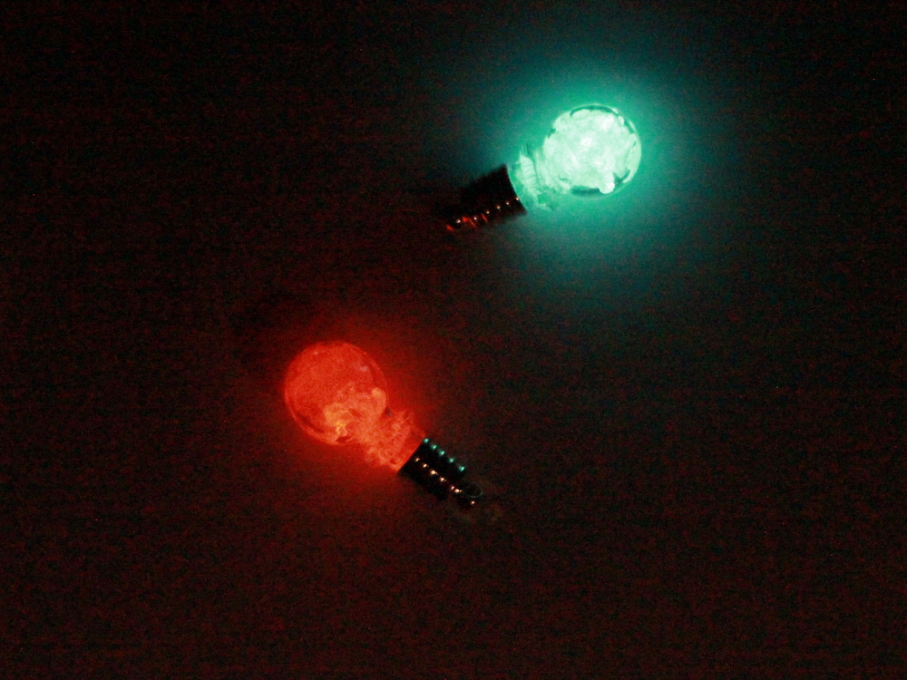
Cyanobacteria fossils - from Berkeley university page

Secondly cyanobacteria can group together with algae to form large layered mat-like structures called stromatolites. Slicing these very thinly reveals tiny cyanobacteria fossils, caught between the layers. There are stromatolytes in Australia that have been alive and growing (very slowly) for millions of years, despite looking faintly uninspiring.

Maybe not quite as impressive as dinosaurs...

While no other bacteria form fossils as nicely preserved as the cyanobacteria, they are capable of leaving behind visible remnants of their existence. The polypeptides of the bacterial cell wall (along with cytoplasm and some secreted lipids) can, under certain conditions, act as nucleation sites for minerals. This eventually leads to the organic cell being replaced by a little mineral cast of the bacteria. There is the possibility for quite a few artifacts with this (artifact being the scientific word for "result caused by the preservation process rather than the bacteria") the most common one being the creation of an artificial nucleus structure. As the bacteria degrades the cytoplasm tends to clump, and the crystallisation of minerals around a cytoplasm clot can in some cases create a structure that looks similar to the structure formed by a nucleus.

Endolithic bacteria that live in rocks can leave behind tiny canals in the rock surface that they bore into. These are a lot harder to find and interpretation is usually helped by the discovery of nearby alive endolithic bacteria. Bacteria have also been found trapped and mumified inside tree resin, a la the Jurassic park mosquito.

Once you start getting larger organisms the bacteria have a brand new niche to exploit. Fossilised bones sometimes show the results of a bacterial infection; while the bacteria themselves are not being preserved their presence is still seen in the fossil record.

Unfortunately although these glimpses are really interesting their also kind of frustrating from a biochemical point of view. They provide clues as to the lifestyles and processes within the bacteria but they are such tiny clues. Past biochemical processes are more often found inside the actual bacteria, by looking at clues in the genome and the genomes of related bacteria, than they are in the remains the bacteria leave behind.

---

Perri, E., & Tucker, M. (2007). Bacterial fossils and microbial dolomite in Triassic stromatolites Geology, 35 (3) DOI: 10.1130/G23354A.1

Westall, F. (2001). Early Archean fossil bacteria and biofilms in hydrothermally-influenced sediments from the Barberton greenstone belt, South Africa Precambrian Research, 106 (1-2), 93-116 DOI: 10.1016/S0301-9268(00)00127-3

Guest Post - Survival of the fittest?

ResearchBlogging.orgI'm very excited about this post, which is a guest post from my sister! She's an undergrad doing biochemistry at Bristol University, and she's currently taking a year working in a research laboratory as part of her degree. She's working with Plasmodium at the moment (which is the little protist that causes malaria) but has sent me a bacteria-related post because she knows me, and she knows my blog and who doesn't love bacteria?

Post - survival of the fittest?

Bacteria have always been very adaptable when it comes to surviving evolutionary stressful situations, such as exposure to antibiotics. Usually some form of mutation will arise leading to the creation of resistant strains of bacteria. These will be selected for via ‘natural selection’ processes and go on to replicate to produce a whole population of resistant bacteria that are able to survive.

However new research looked at a colony of wild type E.coli bacteria in a bioreactor under exposure to increasing levels of the antibiotic norfloxacin and found that no more than 60% of growth was inhibited to maintain a sizable population. The resistance levels of the population as a whole, and of 12 random individuals, was checked every day and it was found that they did not correspond to one another.

The majority of the individual isolates were less resistant than the population as a whole but there was one mutant found that was highly resistant. By isolating the supernatant from the high resistance individual, and conducting gel electrophoresis to separate out the intracellular components, a protein was found that was produced in very high numbers.

This was the enzyme tryptophanase which has the main job of breaking down tryptophan to ammonia, pyruvate and indole. Experiments were done to show that the third molecule, Indole, provided an obvious survival benefit under antibiotic conditions. It upregulated multi-drug efflux pumps which helped in the physical export of the drug and it also had a role in activating various oxidative stress protective mechanisms. The mass production of Indole by the highly resistant mutant allows more vunerable cells in the surrounding area to survive.


a) no antibiotic stress, bacteria naturally produce indole.
b) dead and dying bacteria fail to produce indole
c) mutant appears and supplies indole at a fitness cost to itself.

The resistant bacteria is therefore not selfishly replicating to outgrow the rest of the population but, in helping others to survive, is enduring a fitness cost of its own by mass producing Indole.

This experiment was also carried out using various different antibiotics and the same bacterial altruism was found to exist. The survival of the weaker bacteria does have some advantages as it allows further exploration of mutations that could be even more beneficial to the population. Also, it keeps the opportunity for the bacteria to return to their original state if the stress is temporary, rather than keeping up the energetically wasteful production of antibiotic resistance genes.

So, bacteria working as a team to ensure not just temporary survival but long term advantages for the whole population. Not just survival of the fittest.

---

Lee HH, Molla MN, Cantor CR, & Collins JJ (2010). Bacterial charity work leads to population-wide resistance. Nature, 467 (7311), 82-5 PMID: 20811456

I for one welcome our glowing overlords...

I've already shown you pictures of the glowing bacterial lightbulbs, but in terms of glowing products the cambridge iGEM team is going from strength to strength. To start with the most amazing, they've made an oxygen activated bacterial bubble-lamp:


(It can be found here if the above does not work)

Maybe it's not the most efficient thing to read by, but it's an impressive level of brightness and it turns off relatively (biologically speaking!) fast. It's simply a large measuring cylinder with a load of bacterial broth inside it, and a tube to blow air through. Even shaking lets enough oxygen through to start turning on the light.

Another thing they've been doing is playing around with images on 24-well plates. For those who haven't used them, 24-well plates are usually run on a plate-reader which reads samples from every well, used mostly (in our lab) for overnight assays or (as I should be doing tomorrow) as a glorified spectrometer, measuring a range of absorbances over different wavelengths.

The iGEM team are using them to make nerdy pictures. This is my favourite:

The thing I like about the 24-well-plate pictures is that their different. Painting on plates is awesome buts it's been done before a couple of times, and the glowing pixel-pictures just look new and fresh and exciting.

There's probably going to be a bit of speculation as regards this of the "but how useful is it" type. And rest assured the iGEM team are thinking of that but at the moment I'm happy to just enjoy the fact that we have glowing pixelated space invaders sitting on the bench.

Glowing pixelated space invaders!

This is why I went into science :p

Bacteria that tear themselves apart

This post was chosen as an Editor's Selection for ResearchBlogging.orgBacterial cell division is one of those fairly well studied areas, where time and much study has come forth with a nice standard model. One of the main proteins involved is FtsZ, which seperates one bacteria into two by forming a ring of protein around the middle of the bacteria and tightening it shut as shown below (figure from Nature paper):

Until quite recently it was thought that this was pretty much the only way to get bacteria to divide, until 1999, when the sequences of two Chlamydia species turned out not to contain genes for FtsZ. As Chlamydia are intracellular parasites (which I covered in more detail here) it was at first thought that they might be using some host proteins to complete the cell division, but after the discovery of an FtsZ-less free living archaea, and several more bacteria, it became apparent that the FtsZ-centric model of cell division (shown diagrammatically above) wasn't covering the behaviour of all bacteria.

In the archaeal species (in fact the entire archaeal kingdom Crenarchaea) the cell division was found to be based on a completely different cytoskeletal system. By screening for genes that were turned on at the onset of cell division, a three-gene operon was found to be involved. These genes coded for homologues of eukaryote vesicle trafficking proteins and their regulators, and it was suggested that they formed curved filaments which could pinch off sections of the membrane, forming new archaea. Although the method is similar, the 3D structure of the archaeal proteins is very different to that of FtsZ; the two proteins are not related, but have been coerced into doing the same job.

As well as finding bacteria without FtsZ, it was also discovered that taking a strain of Mycoplasma genitalium and removing the FtsZ didn't stop cell division and in fact showed the same growth kinetics as the wild type. The division mechanism in this case relied on the fact that Mycoplasma move by adhering to a surface and pulling their way along it (in a lab this will be on a glass or plastic surface). To pull their way forward they use a 'terminal organelle', a little protrusion that attaches to the surface and pulls the cell along (figure from the reference).

The diagram above shows Mycoplasma without FtsZ undergoing cell division. You can clearly see not one, but two little terminal organelles at either end of the long stretched cell. What's happening is that in the absence of proper organised proteins to sort out cell division the bacteria has taken matters into its own hands, and sent two terminal organelles determinedly heading off in opposite directions. The bacteria is literally tearing itself apart, splitting into two by ripping in half and letting the membrane close up behind.

It has been suggested that this might be an older method of cell division, used before FtsZ entered the Mycoplasma. It's certainly a lot more brutal than FtsZ-mediated division, and the bacteria has to spend a lot more time in stationary phase recovering from it. As this method only works in bacteria that can attach and hang onto surfaces, it is unlikely to be use by the Chlamydia species (the mechanism for their cell division is still unknown, although some work has been done with L-form bacteria). In the archaeal species it may even be the other way around, that the new filamentous system evolved to be even more efficient that FtsZ in certain species, and so the FtsZ has been dropped entirely.

All of this builds up a picture of just how diverse even simple systems like cell division can be within the bacterial kingdom. And, in my mind at least, is a compelling argument for not just working with model organisms...

---

Erickson, H., & Osawa, M. (2010). MicroCommentary: Cell division without FtsZ - a variety of redundant mechanisms Molecular Microbiology DOI: 10.1111/j.1365-2958.2010.07321.x

---
Follow me on Twitter!

...now in red

Got back from the conference last night, absolutely shattered. I had a great time, and the presentation was really well recieved, everyone seemed to love our coloured bacteria and it was my first time doing a conference presentation to Grad students. I met some new friends, and got the chance to visit Venice on the way back.

While I was away, red happened!

I'm still holding out for the glowing green myself...

To find out more visit the Cambridge iGEM team wiki.

I have a lot of stuff to catch up on, but if anything particularly amazing has happened on the internet while I've been away (other than Bora moving house - which I know about already and wish him the best of luck) drop me a note or stick it in the comments.

Bacterial Lightbulbs

I'm off at a conference in Italy this week, where I'll be doing a presentation about the iGEM work from last year along with some of the follow-up work I've been doing this summer. I'm really looking forward to it, but it does mean that I've got things to concentrate on other than blogging at the moment. I might get the last SGM post out on the weekend but I really need to try and spend that time with my fiancé rather than my science, as I feel I've been neglecting him a bit over the last week to try and get this presentation written.

So in the mean time, have a bacterial lightbulb:

This was made by this years Cambridge iGEM team (their website can be found here) by growing bacteria on agar, mashing up the agar in a little pot and growing again overnight with liquid medium poured over. Some of the resulting bacterial/agar glowing mush was then put into this lightbulb pendant (brought off ebay) to produce a little glowing bacterial lightbulb. Even better the little clumps of agar look a bit like sort of glowing crystals, which gives it a wonderfully 'science'-y effect.

The lightbulb used DNA from Vibrio fischeri, a bacteria that lives symbiotically with squid. The team acquired plasmids containing this gene controlled by the lux operon, and simply cut away the control, meaning the gene is now constitutively expressed (in the presence of oxygen) and glows away happily without any kind of exterior control. Eventually the lightbulb above will start to fade as the bacteria die and require more nutrients, but it's been sitting in the lab for a day now and is still fairly bright.

They've gotten some wonderfully spooky pictures. I don't know if it's just my imagination, or if I'm expecting this of biological systems, but it all looks faintly green to me. Green glowing things are probably the ultimate achievement in science. As well as their current white glow, they are also starting to get some other colours too, ultimately I believe their hoping for green, blue, orange red and yellow.

I'm really impressed with all their work, considering it's the efforts of nine students over a few months. They've got another month to go before they present it at MIT in Boston, which will require frantic powerpoint slides, and (I've been lead to believe) some potential animation work.

See more photos here, visit their wiki here, and follow them on Twitter here.

Bacteria vesicles - SGM series

ResearchBlogging.orgThe SGM autumn conference is now over - thanks to everyone who tweeted it so people like me could catch up on events without actually going. I've just got two more topics of my own little personal blog-conference to go, and this one is going to be on bacterial vesicles rather than secondary metabolism because it suddenly struck me that I don't actually know much about outer membrane vesicles, and this might be a good opportunity to explore them.

So this is the penultimate post in my SGM topic series: Bacterial vesicles.

The first thing to note about them is that they only form in Gram negative bacteria, which have an outer membrane covering a small glycopeptide layer (Gram positive bacteria have no outer membrane and a very large glycopeptide layer). The top layer simple peels off into a little vesicle, taking periplasmic proteins with it, as shown below (diagram from the reference):

The mechanism for vesicle formation is largely unknown, but it is found in both pathogenic and non-pathogenic strains of bacteria, and used for several different purposes. In pathogenic bacteria the vesicles often contain virulence factors, which can destroy or damage host cells. in the wild, they may also bind to or destroy other bacteria. In less-virulent strains they have been shown to act as a method of removing misformed or unwanted proteins from the periplasmic space (the space between the two membranes). They can also play a part in antibiotic resistance, it's not yet certain how but my guess is that they pump the antibiotic into the periplasmic space then vesicle it off to stop it just diffusing back in again.

When first discovered, the vesicles were thought to be a by-product of bacterial death, after all, why else would little bits of membrane with bacterial proteins inside be found floating around a large colony? However work done on pathogenic bacteria (which get more funding) and biopsies of infected tissues showed the vesicles playing an important part in infection. They are produced during the stationary phase of growth, the same period when bacteria start to produce most of their virulence factors and secondary metabolites. In an infected organism, this phase is after the bacteria has set off an inflammation reaction, and once it has multiplied in the site of infection.

Factors that affect the formation of vesicles include oxygen stress, the availability of iron (finding a regular iron source inside human bodies is a regular problem for bacteria) and the composition of the outer membrane (suggesting that at least some of the mechanism is mechanical). It is a ubiquitous process carried out across a range of Gram negative species.

As well as being used offensively, some vesicles were also shown to carry DNA between bacteria, although it's not at all clear how, or how the DNA gets into the periplasm in the first place. P. aeruginosa are also capible of transferring antibiotic resistant enzmyes between bacterial cell using the vesicles. This is not totally an act of complete altruism, as P. aeruginosa carries out much of it's infectious cycle as a biofilm, which requires lots of cells to form.

As I said, the actually mechanism for the formation of the vesicles is not yet established, so there's probably quite a lot of work to do with imaging their formation, genetics to find out any genes involved, and a mixture of genetics and protein work to discover more about what goes inside the vesicles. It looks like an interesting area of research, with the potential for some quite amazing imagery-work, and I look forward to reading more about it.

---

Kuehn MJ, & Kesty NC (2005). Bacterial outer membrane vesicles and the host-pathogen interaction. Genes & development, 19 (22), 2645-55 PMID: 16291643

Surviving in the stomach - SGM series

The fifth post now in the SGM series, and this one focuses on Acid Stress. As I covered in a previous post, bacteria are able to survive in a number of adverse conditions, so this post will focus on just one of them, high acidity. Particularly the high acidity within the human stomach.

There are many different bacteria with acid stress survival techniques, all bacteria that ever visit the stomach need to be able to cope with the low pH. Listeria monocytogenes is a bacteria that anyone whose every looked into pregnancy will know about, as although it does not often cause human dieases it can have potentially fatal effects on an unborn fetus. The bacteria are found in un-pasteurized cheeses and dairy produce, and apparently one of the reasons for this is that the food surrounding them helps to protect them against the acidity of the stomach.

Soft cheese - now with added Listeria!

There are various different ways that a bacterial cell can survive acid stress (which is distinct from surviving in constant acidic conditions). Regulatory systems for acid stress have been found, such as the two component EvgS/EvgA system in E. coli, which turn on a large number of acid-stress related genes when sensing low pH in the surrounding environment. In Bacillus cereus, which hides out in warm rice (and is the reason you have to re-heat takeaways to piping hot the next day at work) acid stress conditions were shown to induce a number of typical bacteria cellular responses - two component systems, alternative sigma factors (which lead to changes in the proteins being produced) and excess proteases and chaperones to get ensure more correctly folded proteins (harder in an acidic environment).

With the Listeria however, it was discovered that they were actually getting a lot of help from their surrounding food. Certain amino-acids, such as glutamate, are used by the bacteria to neutralize stomach acid and it turns out that cheese is quite high in glutamate. Also, a fair amount of acidity is used in the cheese making process, which helps the bacteria practice at their acid-resistance; essentially any bacteria in contaminated cheese will have already run an acidity gauntlet which stands it in good stead for the experience it will get in your stomach.

This work is also interesting as it shows that it's not just the amount of consumed bacteria that can lead to stomach poisoning, it's also dependent on the surrounding nutritional environment of your stomach. Eating Listeria in non-glutamate containing food is less likely to lead to an infection, unless you also like tomato juice (very high in glutamate) and drink that at the same time. As well as being interesting from a scientific view this is also very useful diet advice for people susceptible to certain bacterial infections. An understanding of how bacteria work within specific metabolic environments will allow better dietary advice about foods to keep away from.

---
There is no paper for this, so I'm working mostly off the press release, which can be read here. As I didn't attend the talk at the SGM conference, I'd welcome any corrections of any facts I may have either omitted or misinterpreted. Having read the abstract I'm kindof gutted I didn't get to this talk, it looks fascinating.
---

Follow me on Twitter!

Studying Streptococcus - SGM series

ResearchBlogging.orgThe fourth post now in the SGM series, and this one focuses on Streptococci. Streptococci are a genus of spherical Gram-positive bacteria containing both pathogenic and harmless strains, including the flesh-eating bacteria (which cause the delightfully named necrotizing fasciitis) as well as bacteria responsible for making swiss cheese. Commensally they are found on many parts of the human body, including the mouth, skin, intestine, and upper respiratory tract .

Streptococcus - growth and division leads to long chains of bacteria (image from lenntech)

It's quite a broad topic which allowed plenty of speakers to address their favourite issues with these bugs, but as well as discussions of the virulence factors, biofilm properties and various different intracellular survival properties of the Streptococcus there were also some talks covering new research mechanisms. Rather than focusing on the properties of the bacteria, these talks were about new methods used to study them.

The one that jumped out at me the most was about using Bioluminescent imaging to track a Strep infection. This appealed to me because the iGEM team next door are working on Bioluminescence so it's a word I've heard a lot over the last eight weeks. By adding bioluminescent bacteria to a mouse model, the course of the infection can be tracked over several weeks (using small animal imagine machines it can be tracked in the same mouse). This provides a far better understanding of the pathogenesis of the bacteria; how it spreads through the body and at what point it is most infectious.

The process of using bioluminescence to track diseases (image from the reference).

Using luminescence to study disease progressions isn't a new idea, but the use of whole animal scanning mechanisms now means that fewer animals have to be sacrificed in order for the study to done. The luminescent tissue does not have to be extracted, and the more natural disease progression can be followed.

Other methods explored included the by-now predictable whole genome study analysis to organise the different types and virulence levels of a Streptococcus suis which leads to meningitis in piglets. Comparative genome hybridization studies allow many genomes to be compared at once, giving a better idea of the differences and similarities between them. This helps to separate the strains into serotypes (different groups), and to compare the differences that lead to virulence. Genome comparison work was also being done for Streptococcus equi species which cause infections in horses.

In other news (pretend that was a smooth transition!) the latest Carnival of Molecular Biology is out over at Thoughtomics. There are some brilliant articles covering the intra-cellular happenings of organisms from bacteria to frogs to Tibetans. If you've ever wondered about noisy bacteria, zombie enzymes or what micro-RNA is, go take a look and visit the submissions.

---


Timothy C. Doyle, Stacy M. Burns, Christopher H. Contag (2004). In vivo bioluminescence imaging for integrated studies of infection Cellular Microbiology, 303-317 DOI: 10.1111/j.1462-5822.2004.00378.x

Xtreme bacteria! - SGM series

This is the third post in the SGM series and this one is about Extremophiles. There were lots of talks in this topic, so instead of a serious research-analysis post, this will just be a quick run-down of some of the more interesting bacteria that were covered:

Award for : Survival under extreme acidity
This award is presented to the Natranaerobiales species that live in sun-heated salt lakes in the Middle East and Africa. These can grow at conditions of Ph 3.7 and also manage to survive at 66 degrees Celsius, by using specially modified proteins to carry out vital cellular tasks.

Award for: Coldest temperature - thermophiles
Theremophiles are bacteria that are found only in very hot temperatures, so researchers were rather surprised when an experimental heating up of arctic ice to 50 degrees stimulated a range of dormant thermophiles to spring to life. What the bacteria were doing there, what kind of global warming event they were waiting for, and why researchers were heating up arctic ice to 50 degrees anyway remains to be discovered...

Award for: Resistance to radiation
This award collected on behalf of all radiation resistant bacteria by Deinococcus radiodurans. Recent work in these bacteria revealed that there is a mechanistic link between resistance to radiation, protein protection and (rather weirdly) manganese accumulation. Apparently surplus manganese in cells can help against reactive oxygen species.

Award for: Survival at greatest pressure range
The deep-sea bacteria Photobacterium profundum is not only able to survive at 15 degrees C, but also lives at a pressure of 28MPa (atmospheric pressure being 0.1MPa). Any eukaryotes living at that pressure tend to unfortunately explode when brought to the surface, but these bacteria are able to survive fine at normal pressures. (Scientific aside - their cell surface lipopolysaccharide composition is thought to help with the cold survival. Mutants that are cold sensitive have a lower concentration of smooth LPS than the cold-adapted parents).

Award for: Best attempt at mimicking a B-list Sci-Fi alien
I'm not sure whether this should be given to the bacteria, or the people who wrote the abstract, but to give you a clue the talk is titled "Dark life in the fracture labyrinth of deep hard rock". Bacteria have been found in rock fissures living in complete darkness with hardly any organic minerals, and using hydrogen as an energy source. Or, in the words of the abstract, "An extreme ecosystem that survives in total darkness feeding by hydrogen from the interior of our planet". If these things weren't tiny blobs that would be a movie already.

Honorable mention: Extreme fungi
This was a microbiology conference, not a bacteriology one, which means that it also covered fungi, algae and protists. Fungi have been found that live at high salt concentrations, both high and low temperatures, acidic and basic conditions, high hydrostatic pressure, high ionizing radiation and in extremely toxic environments.

Honorable mention: The virus's of Haloquadratum
These little guys are getting an honorable mention just because they seem to be the only virus's mentioned in the entire talk. They infect the bacteria Haloquadratum and are thought to have played a role in the development of the bacterium's resistance to high salt levels.

That's all the awards for now! Just four more of the SGM series to go, I'm enjoying writing them, even if the "one post every two days" hasn't quite happened.

How bacteria die - SGM series

ResearchBlogging.orgThis is the second post of my SGM conference series and the topic is Microbial Death. I was very interested in this one as a topic, because the mechanisms that lead to bacterial death aren't something I've covered so much. It's generally assumed that antibiotics screw up whatever they target such that the bacteria can no longer survive, and when they aren't around the bacteria just keep dividing.

There were two talks concerning antibiotics in bacterial death, the first addressing a theory that's been bandied about for a while (and which I've already written about) that antibiotics don't really kill the cell by acting on their target. Instead, they just lead to sufficient damage to set off a series of death events within the bacteria themselves, a common pathway for bacterial cell destruction (first reference).

I think if I could have chosen any one talk to watch it would have been that talk, actually given by Kohanski whose been working on the stuff. I think there would have been some interesting questions as well, as this is somewhat controversial research.

The other antibiotics talk covered something I'd never heard about; the ability of some antibiotics in certain cases to prevent bacterial death. Work done on Microbacterium turberculosis - which causes TB and a related strain (Microbacterium bovis) showed that when in stationary phase (i.e the bacteria were not growing and dividing) the addition of antibiotics that usually kill only growing cells helped to aid cell survival. Antibiotics that targeted both growing and non growing cells did not have this effect. The reason for this is not clear, however comparing transcriptomes between cells both with and without antibiotics showed a difference in protein production on addition of antibiotics. These antibiotics are in someway helping to turn on genes for survival, which are keeping the stationary phase bacteria alive.

Another interesting talk was about the regulation of mutagenesis in bacteria, another idea I love. It's based on the observation that as bacteria start to get stressed they go into a sort of massive meltdown, leading to lots of genetic mutations being generated. It's been suggested that rather than this being a side-effect of the surrounding stress, this is actually a deliberate ploy by the bacteria to give themselves a last ditch attempt at getting out of a stressful situation.

Unlike multicellular organisms, bacteria have no surrounding restraints on their mutation rate - with the exception of bacteria in aggregates the only thing a bacteria will harm by changing it's DNA is itself. This gives bacteria a lot more genetic plasticity. Added to this, changing DNA is one of the main ways bacteria go about improving themselves, and adapting to new conditions. Changing the DNA by wholescale random mutagenesis is a bit extreme, but if you're in a stressful situation anyway it might be worth a shot.

Studies for this have mostly been done on E. coli, usually a lab strain, so I'm not sure how much they translate into bacteria in the wild, which might be better adapted at coping with stress situations, or, given that experimental bacteria are in a privileged nutritional environment, it might just be too risky for wild bacteria to start messing around with their genome. Also there's no concrete mechanism been found for it yet, so increased mutagenesis producing different phenotypes in times of stress may just be a happy byproduct of the usual genetic craziness that goes on when a cell dies.

These two theories both don't have as much supporting science as they could do, but they are new ideas which are still being worked on, and I really like them both.

---
Kohanski MA, Dwyer DJ, Hayete B, Lawrence CA, & Collins JJ (2007). A common mechanism of cellular death induced by bactericidal antibiotics. Cell, 130 (5), 797-810 PMID: 17803904

Ivana Bjedov, Olivier Tenaillon, Bénédicte Gérard, Valeria Souza, Erick Denamur, Miroslav Radman, François Taddei, Ivan Matic (2003). Stress-Induced Mutagenesis in Bacteria Science, 1404-1409 DOI: 10.1126/science.1082240

---
Follow me on Twitter!