A
friend of mine, who actually reads my blog occasionally, was very interested in the idea of DNA sequencing, fascinated by the thought that DNA could just be created in companies and then shipped out when needed. He mentioned I should write a post on DNA sequencing.
I
thought about it, and realised, with a daunting sense of dread, that actually I would have to do quite a bit of research before being able to write coherently about DNA sequencing. I know the
general idea, but not enough to explain to someone who doesn't already have quite a good idea of whats going on. Luckily there already is a very clear and comprehensive
explanation of it over at Genetic Interference:
Part OnePart TwoSo I'll just add to the story a little by describing things from the point of view of me...the scientist actually ordering the DNA.
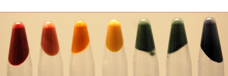
First I need to find out what I want. This requires a literature search. For
example, when I started looking for my pigment colours, I went on a quick trawl through
PubMed, looking for any genes that had been shown to produce colour in E.
coli. The
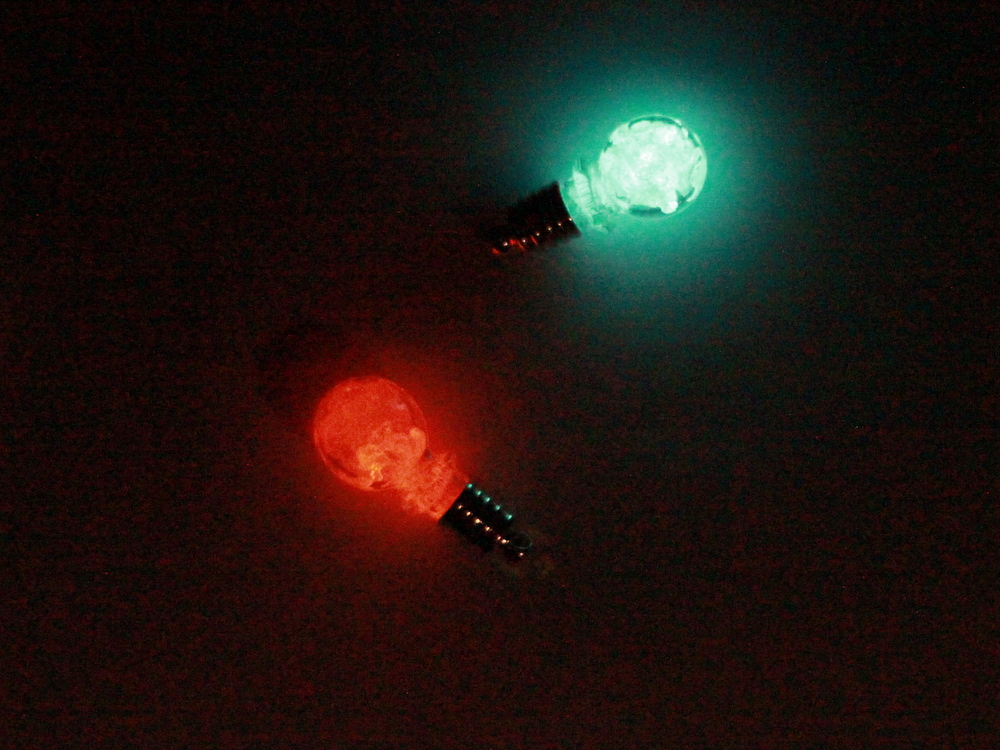
vio gene shown in
this post is just one example, at the moment I also have a brown pigment, and (hopefully soon) two genes that make green and red as well.
The next stage is to find the actual DNA sequence. Usually it's in the PubMed paper, or in
NCBI - which has a huge database of all proteins that people have registered. If it's very new research, you might have to email or phone the researchers. Once you have the DNA it's a good idea to double-check it as well...compare to homologous proteins or ones with similar domains. I used the
MUSCLE comparison tool for this,
simply because it's the one I'm most used to.
Once you're certain you have the right sequence for what you want, you contact a DNA synthesis company. Prices vary... as far as I can work out it varies from 20 (if you're REALLY lucky) to 50p per base pair. A smallish gene is usually about 1kilo-
base pair(
kb) just to give an idea of the scale of things. And the price tends to just up once you get over 1
kb as well. The
vio gene which we are getting for free is about 6
kb long.
Then you send your sequence off to get made! You can have various options for synthesis (as I am just discovering). The
codons (AAA,
GGG etc) can be optimised for your organism - in the case of more than one
codon (the three bases) coding for one amino acid, different organisms will prefer
to use different
codons. You can get restriction sites removed and added (for cutting and pasting DNA parts), and extra parts added to the gene, such as an area for the beginning of protein coding, or a degradation tag, which will cause the end product protein to break down (very useful if it's a long-living protein you want to get rid of quickly).
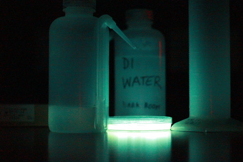
Larger genes get sent in bacteria, on little loops of DNA called plasmids to keep them replicating inside the bacteria. You grow your culture up, and then can extract your precious DNA from them. Small bits of DNA, like primers, just
arrive as naked DNA, inside a little plastic vial, and can be made up to solution with water.
After synthesis, it's a good idea to get them sequenced as well... just to check you have the right stuff.