Field of Science

Showing posts with label antibiotics. Show all posts

Coloured bacteria vs. Angry birds

For those who didn't catch the news when it first came out, the iGEM project I carried out in the summer of '09 has been nominated for a Design Award! The Brit Insurance Design of the Year 2011 to be precise.

I'm very excited about it, even though I don't really feel like a designer (I don't own a singlething made by Apple). We're up against some pretty intense competition, such as the Angry Birds phone game, Rock band 3 and quite a few phone apps. Still, it's pretty awesome to be nominated, and I'm looking forward to seeing all the rest of the project displays.

Our two wonderful designer-friends James King and Daisy Ginsberg have put together a great video explaining the project. It's got some great animation, and clips from a radio interview with one of my fellow lab rats, and even some pictures of the infamous Orange Liberation Front...

E. chromi from Alexandra Daisy Ginsberg on Vimeo.

Targeting dormant bacteria

ResearchBlogging.orgAntibiotics are effective against bacteria because they target and knock out specific functions that are vital for bacterial survival. As most bacterial infections involve rapid growth and division of the invading bacteria, many commercial antibiotics currently target metabolically active cells, by blocking enzymes needed for growth, reproduction, or cell wall synthesis. While these will kill acute bacterial infections they are often far less effective against dormant bacteria in longer-term persistent infections.

Rather than targeting metabolic enzymes, the current strategies being explored to combat dormant bacteria target either the membrane, or membrane bound proteins. Both of these approaches destabilise the bacterial membrane and help to break the cell apart and can act against processes such as energy synthesis which occur in both active and dormant cells.

a=targeting important metabolic proteins in the membrane. b=targeting the actual cell-membrane. Picture is copywrite me :p

In eukaryotic cells, such as the cells of plants and animals, the enzymes that create energy for the cell are kept safely hidden away in specialised intracellular compartments, such as mitochondria. As energy production requires an ion gradient across a membrane, these compartments all have sets of internal membranes. Bacteria however do not have this luxury, and instead have all their metabolic enzymes in the outer cell membrane, as this is the only membrane they have. Inhibitors of energy metabolism can therefore bind directly to target enzymes in the membrane involved in the production of energy. This can be highly effective against cells whose interior is hard to get into, such as Mycobacterium tuberculosis which lurks inside tuberculosis granulomas. Even in the absence of growth, cells still require a minimal energy input to survive, so blocking off these enzymes kills both dormant and active cells.

Drug developed to help combat TB by attacking cell membrane metabolic enzymes. This drug is currently in stage three clinical trials.

The membrane-targeting drugs act directly on the lipid bilayer that surrounds the bacterial cell, breaking it up and destroying the bacterial cellular integrity. Although human cells are also surrounded by lipid bilayers they have fewer negatively charged phosopholipids and also contain cholesterol (not present in bacterial membranes) allowing membrane-targeted drugs to be specific for human pathogens rather than killing surrounding human cells. The drugs that are used to attack the cell wall can vary hugely in size and structure but they all share one common property; they are highly lipophilic (i.e they are attracted to lipids). This allows them to interact with the cell membrane and break it apart.
Lipophilic drug capible of targeting bacterial cell membranes

There’s something about those molecular diagrams of drugs that I love. I think it’s my biochemical background. I’m never totally happy with a schematic until I can see how the chemicals are interacting on a molecular scale.

As well as being useful against dormant bacteria these new antimicrobials show promise as strategies for dealing with arising antibiotic resistance. Bacteria can evolve to cope with as many challenges as are thrown at them, but hopefully it should take them a little longer learn to survive entirely without a cell wall…

Although there are some that can do that already.

---
Hurdle JG, O'Neill AJ, Chopra I, & Lee RE (2011). Targeting bacterial membrane function: an underexploited mechanism for treating persistent infections. Nature reviews. Microbiology, 9 (1), 62-75 PMID: 21164535

---
Follow me on Twitter!

Antibiotics and gut bacteria

This post was chosen as an Editor's Selection for ResearchBlogging.org
All microbiologists end up writing about gut bacteria at some point. It is the way of things. Disease of the Week is currently doing a whole series on it, and a few weeks ago I covered the interaction of the immune system with gut bacteria (here). However a recent paper came out in Microbiology Today concerning the affect of antibiotics on gut bacteria, which is a topic that I both find interesting and have had some actual experience with.

I've taken antibiotics a few times, and each time I've found that despite the many positive effects it has (i.e I don't die of septicaemia) being on antibiotics tends to make my stomach fairly unhappy. I usually have a good relationship with my stomach - I feed it regularly with plenty of food and in turn it is generally quite accepting of the fact that I don't always wash my hands as much as I should, and occasionally eat things that might be past their sell-by date, or heated up food that isn't exactly the "piping hot" recommended by the label.

Antibiotics however are usually designed to kill off bacteria, and unless the antibiotic in question is very specific that often includes your commensal gut bacteria; the 'friendly' bacteria that yoghurt companies keep mentioning, that hangs around in your gut and prevents other bacteria invading. In the short term this leads too the occasional unhappy stomach but in the long term it can have a more sinister effect, by encouraging antibiotic resistance genes to develop and spread amongst the normal bacteria in your gut.

The diagram below shows a stylised (and not to scale) diagram of what happens to your gut bacteria after a dose of antibiotics. The antibiotic resistant bacteria (in purple) are suddenly at an advantage and can proliferate. As time goes by, the other natural bacteria gradually return to your gut (which is not after all the most sterile of environments) but there will be a higher concentration of antibiotic-resistant bacteria around.

Diagram taken from the reference (below)

It's important to remember that these antibiotic-bacteria are not dangerous in themselves. Unless they move out of the gut for any reason they will remain totally harmless. However they also contain the genes for antibiotic resistance, and if another bacteria manages to survive and get into the gut, they can pass those genes on. As every human carries different gut bacteria, and will take different antibiotics in the course of their lifetime, this study cannot really make any hard and fast rules about what the effect of this might be on any one individual, but it does make the point that many people will be carrying within them a large proportion of bacteria that may have developed a novel form of antibiotic resistance.

It's not always terrible, in some cases the antibiotic resistant bacteria are grossly unfit, and are immediately out-competed as soon as the antibiotic goes away. Those that do survive both the antibiotic and the returning bacteria however, can hang around for a long time, and antibiotic resistance was seen in gut bacteria up to four years after antibiotic treatment had finished. And the intestines are such a wonderful environment for bacteria to share DNA in; there are abundant nutrients, everything is all moist and warm and there are a lot of bacteria in very close contact with each other.

If you have taken antibiotics it's not worth loosing sleep over, but it's something hospitals are starting to be more and more aware of.

---


Jernberg C, Löfmark S, Edlund C, & Jansson JK (2010). Long-term impacts of antibiotic exposure on the human intestinal microbiota. Microbiology (Reading, England), 156 (Pt 11), 3216-23 PMID: 20705661

Chopping bits out of the genome

ResearchBlogging.orgGenerally bacteria genomes tend to be fairly minimal in the amount you can remove from them. Unlike eukaryotes, which can have whole swathes of genome that codes for very little, bacteria, with their limited space for a chromosome, need every gene they can get. They just don't have the space for unnecessary genes.

Streptomyces bacteria, however, have bigger genomes and the luxary to invest in genes which are not strictly necessary for bacterial survival. These are called Secondary metabolite genes (as opposed to the necessary primary metabolites) and they code for genes that form an arsenal of weapons for the Streptomyces to deploy. Most Strep are soil-based, and they need the ability to produce secondary metabolites (such as antibiotics) to fight off invading bacteria, and clear terratory to expand their growth into.

What has recently been done (very ingeniously) is to remove the secondary metabolism genes from the bacterial species Streptomyces avermitilis creating essentially an 'empty' strep bacteria, that can grow and divide but not produce any of the secretory substances that strep are known for. The researchers managed to cut an entire 1.5Mb of DNA right out of the genome - helped by the fact that all the secondary metabolite genes cluster together on one side of the chromosome.

They did this using a common molbio technique, the cre-lox system. "Lox" is an area of DNA and "cre" is the protein that very specifically cuts DNA at the Lox site - it acts like a pair of scissors. They put a Lox site on either end of the DNA that codes for secondary metabolites (using a techinque called recombination in order to attach the lox into the chromosome) along with the DNA for the Cre protein under an inducible promoter. Once the bacteria had grown, they activated Cre production, which then cut the unwanted DNA out of the bacteria. This technique was amazingly successful and is shown diagramatically below (picture from the reference):


The avimitilis now contains no secondary metabolites at all, which makes a wonderful 'empty' system to use for studying how secondary metabolites are made. Genes from other bacteria, or even some of the removed genes, can be added back in, piece at a time, to see how much of the gene is necessary for metabolite production, and which regulatory pathways are the most important. Creating an empty cell also has potential implications for biotechnology, after all when trying to produce antibiotics you want the cell working as hard as possible just to produce your product, not wasting time and resources on other metabolites!

I'm really impressed at this kind of large scale synthetic-biology. It may be an area I end up going into in the near future...

---

Komatsu M, Uchiyama T, Omura S, Cane DE, & Ikeda H (2010). Genome-minimized Streptomyces host for the heterologous expression of secondary metabolism. Proceedings of the National Academy of Sciences of the United States of America, 107 (6), 2646-51 PMID: 20133795

How bacteria die - SGM series

ResearchBlogging.orgThis is the second post of my SGM conference series and the topic is Microbial Death. I was very interested in this one as a topic, because the mechanisms that lead to bacterial death aren't something I've covered so much. It's generally assumed that antibiotics screw up whatever they target such that the bacteria can no longer survive, and when they aren't around the bacteria just keep dividing.

There were two talks concerning antibiotics in bacterial death, the first addressing a theory that's been bandied about for a while (and which I've already written about) that antibiotics don't really kill the cell by acting on their target. Instead, they just lead to sufficient damage to set off a series of death events within the bacteria themselves, a common pathway for bacterial cell destruction (first reference).

I think if I could have chosen any one talk to watch it would have been that talk, actually given by Kohanski whose been working on the stuff. I think there would have been some interesting questions as well, as this is somewhat controversial research.

The other antibiotics talk covered something I'd never heard about; the ability of some antibiotics in certain cases to prevent bacterial death. Work done on Microbacterium turberculosis - which causes TB and a related strain (Microbacterium bovis) showed that when in stationary phase (i.e the bacteria were not growing and dividing) the addition of antibiotics that usually kill only growing cells helped to aid cell survival. Antibiotics that targeted both growing and non growing cells did not have this effect. The reason for this is not clear, however comparing transcriptomes between cells both with and without antibiotics showed a difference in protein production on addition of antibiotics. These antibiotics are in someway helping to turn on genes for survival, which are keeping the stationary phase bacteria alive.

Another interesting talk was about the regulation of mutagenesis in bacteria, another idea I love. It's based on the observation that as bacteria start to get stressed they go into a sort of massive meltdown, leading to lots of genetic mutations being generated. It's been suggested that rather than this being a side-effect of the surrounding stress, this is actually a deliberate ploy by the bacteria to give themselves a last ditch attempt at getting out of a stressful situation.

Unlike multicellular organisms, bacteria have no surrounding restraints on their mutation rate - with the exception of bacteria in aggregates the only thing a bacteria will harm by changing it's DNA is itself. This gives bacteria a lot more genetic plasticity. Added to this, changing DNA is one of the main ways bacteria go about improving themselves, and adapting to new conditions. Changing the DNA by wholescale random mutagenesis is a bit extreme, but if you're in a stressful situation anyway it might be worth a shot.

Studies for this have mostly been done on E. coli, usually a lab strain, so I'm not sure how much they translate into bacteria in the wild, which might be better adapted at coping with stress situations, or, given that experimental bacteria are in a privileged nutritional environment, it might just be too risky for wild bacteria to start messing around with their genome. Also there's no concrete mechanism been found for it yet, so increased mutagenesis producing different phenotypes in times of stress may just be a happy byproduct of the usual genetic craziness that goes on when a cell dies.

These two theories both don't have as much supporting science as they could do, but they are new ideas which are still being worked on, and I really like them both.

---
Kohanski MA, Dwyer DJ, Hayete B, Lawrence CA, & Collins JJ (2007). A common mechanism of cellular death induced by bactericidal antibiotics. Cell, 130 (5), 797-810 PMID: 17803904

Ivana Bjedov, Olivier Tenaillon, Bénédicte Gérard, Valeria Souza, Erick Denamur, Miroslav Radman, François Taddei, Ivan Matic (2003). Stress-Induced Mutagenesis in Bacteria Science, 1404-1409 DOI: 10.1126/science.1082240

---
Follow me on Twitter!

Nanofibre paint that kills MRSA

This post was chosen as an Editor's Selection for ResearchBlogging.orgMRSA, the antibiotic resistant form of Staphylococcus aureus is a major problem in hospitals. The antibiotic resistance makes it hard to erradicate, not just from patients, but in the surounding environment, on surfaces, on medical equipment, on the walls of the hospital. In order to minimise the numbers of dangerous bacteria found in hospital surroundings, quite a lot of research has gone into creating antibacterial coverings or coatings that would reduce the number of bacteria p. Currently however, many of these coating substaces work by either using biocides (such as silver) or releasing antibiotics and antimicrobials, which doesn't work on bacteria that have gained resistance.


Scanning electron microscope picture of clusters of Staph aureus

However medical scientists aren't the only ones trying to kill bacteria, virus's known as bacteriophages are also interested in breaking open bacterial cells and they do it using a cocktail of different enzymes to break open the cell wall. Many of these enzymes feature a two-domain structure with bacteria-specific cell wall targeting and catalytic domains. The enzymes Lst was found to be particularly good at breaking apart Staph aureus cell walls with one end of the enzyme (C terminus) recognising and binding to the bacterial cell wall while the other end (the N terminus) breaks the protein bridges between the sugar componants of the peptidoglycan layer, which is a major componant of the cell wall.

Schematic of the cell wall

Work from the Rensselaer’s Center for Biotechnology has been looking at incorporating these enzymes into nanofibres to create stable bactericidal paint films. The molecular-level curvature of carbon nanotubes stabilizes a wide range of enzymes and the lab was able to successfully create Lst-containing nanocomposite films which achieved >99.9% killing of MRSA upon contact within 2 h. They also explored incorporating these into a latex paint, which retained the bactericidal properties of the nanofibres. This paint could theoretically be spread over hospital surfaces to reduce the numbers of Staph aureus within the hospital environment. Incorporating this enzyme into the nanofibres (rather than directly mixing with the latex) gives added stability and helps the enzyme stay within the coating for longer. Films which were stored dry at room temperature showed >99% bactericidal activity against S. aureus after 30 days.

It remains to be seen how effective this technique will be outside of a laboratory setting but at the moment it looks like a highly promising step to help reduce the incidence of a dangerous pathogen. The speed and likelihood of resistance also remains to be seen, but it's heartening that bacteriophages have been using these enzymes against the bacteria for far longer than we've been using antibiotics. This is unlikely to be any kind of magical anti-MRSA cure, but it could certainly be very useful in helping to reduce the incidence of the disease.

---
Pangule RC, Brooks SJ, Dinu CZ, Bale SS, Salmon SL, Zhu G, Metzger DW, Kane RS, & Dordick JS (2010). Antistaphylococcal nanocomposite films based on enzyme-nanotube conjugates. ACS nano, 4 (7), 3993-4000 PMID: 20604574
---

Follow me on Twitter!

Models for the evolution of bacterial resistance

This post was chosen as an Editor's Selection for ResearchBlogging.orgI wrote about Quorum Sensing recently, the ability of bacteria to communicate with each other via small molecules which they can both excrete and sense. A lot of the research done on quorum sensing aims to find ways to block the system, as it is one of the main communication methods used to switch on virulence genes, and other genes which make the bacteria more infectious, and more likely to cause harm.

Blocking quorum sensing would not in theory kill the bacteria, but it would give more time for the bodies immune system to recognize and destroy the bacteria. This is starting to be a more common approach in the design of anti-microbials, and in virus vaccines. A slower and less virulent form of both virus's and bacteria can sometimes be dealt with easily enough by the body's own immune system as it has more time to respond to the invading challenge.

There is also an evolutionary reason for the effectiveness of quorum sensing disruption over conventional antibiotics. Antibiotics kill off any bacteria that aren't resistant, creating a highly competitive growth environment with a large selection bias against non-resistant bacteria. Disrupting the quorum sensing system on the other hand should mean that the bacteria are still growing in a non-selecting environment, right up to the point where the body's defense system kills them off. There is less evolutionary pressure on them to develop resistance.

A paper in PLoS (reference below) looked at developing a method for how (and whether) resistance towards quorum sensing disruption might occur. They chose to base their model on the effects of altering the genes involved in quorum sensing, rather than other methods for quorum sensing disruption, such as blocking or destroying the little molecules used for signalling and one of the first things they found was that there are a large number of variations in these genes, despite the fact that many of the actual signalling molecules are fairly similar.

Lots of gene variation means bad news for gene-based disruption as there is potential for the bacteria to share these genes among themselves, and thus potentially end up with a combination that renders the gene-blocking anti-bacterial invalid. And swopping genes is one of the things bacteria do best.
Figure above shows two bacteria connecting together via a long thread-like molecule called a 'pilus'. The genes do not actually travel through the pilus, but the pilus pulls both bacteria close together allowing the gene (in the form of a plasmid) to be passed between them.

Another thing that was examined was the effect of quorum sensing disruption on fitness. The idea that resistance to quorum sensing (which will henceforth be called QS as I can't be bothered to keep typing it out) will take more time to develop is based heavily on the idea that tampering with the QS does not affect the general health of the bacteria. Sure it prevents bacteria from communicating, but each individual bacteria is no less likely to thrive, and therefore there is less selection pressure.

However almost all of these experiments (certainly those in academic labs) have been done in conditions of nice safe uber-rich bacterial growth media. Even slightly shaken bacteria may thrive fine on nutrient rich agar. Inside an actual living organism though (the normal living place of pathogenic bacteria) disrupting the QS system has major implications. Not just in the fact that non QS-able organisms are often outcompeted by other bacteria, but also because some QS-controlled proteins seem (under certain conditions) to be vital for bacterial growth. If disruption of QS is leading to bacterial death then this puts a massive evolutionary pressure on bacteria to evolve resistance, similar to the pressure to evolve antibiotic resistance.

Interestingly (if slightly off topic) it is vaguely suggested that the necessity of QS for growth might help to prevent 'cheaters' within a bacterial population; bacteria that take up all the benefits of it's QS-ing neighbors (i.e biofilm formation, increased virulence etc) without having to expend energy into the QS system.

In view of this data it looks as if, despite it's undoubtable advantages as an antimicrobial, QS-disrupting systems will inevitably succumb to bacterial resistance. In the presence of a selective pressure favoring it resistance will always develop. There is no golden bullet for antibacterial strategies, just a lot of little silver bullets that eventually loose their power.

So as not to end this on a depressing note, it is worth mentioning that the fact that there is a lot of research going into alternative (i.e not antibiotics) antimicrobials is encouraging. Phage therapy and QS disruption will probably never replace antibiotic therapy, but it is worth having as many different strategies as possible to deal with the threat of bacterial infection.

---
Defoirdt T, Boon N, & Bossier P (2010). Can bacteria evolve resistance to quorum sensing disruption? PLoS pathogens, 6 (7) PMID: 20628566

---
Follow me on Twitter!

Quorum Sensing and Biofilms

This post was chosen as an Editor's Selection for ResearchBlogging.orgAlthough bacteria live as isolated cells, they are constantly communicating with surrounding bacteria, particularly those of the same species, which can often band together to form large groups of bacteria surrounded by a sticky mesh. These are known as biofilms (which I cover in more detail here). One of the main ways that bacteria communicate with each other in order to organise structures like this is by quorum sensing.

Quorum sensing uses small molecules that bacteria can both excrete and sense. When enough bacteria are in one place then the surrounding concentration of these small molecules reaches a critical level and can activate the genes for a variety of different responses including luminescence, virulence (in pathogenic or disease-causing bacteria) and the formation of biofilms:
A recent paper (reference below) looked at a range of different types of biofilm and quorum sensing interactions, in order to explore the different environmental pressures that shaped the differences in these systems. They found that although many species formed biofilms when bany cells joined together some species stopped forming biofilms when they reached a certain cell density. Biofilms are carefully controlled by bacteria, they do not just start growing when a certain number of bacterial cells gather together and then never stop.

Using models of mostly infectious biofilm-forming bacteria (such as Vibrio cholerae which causes cholera) they found that as well as helping to bind the cells together and resist man-made antibiotics (which cannot penetrate the biofilm) the biofilm was also a defense against competing bacteria (and may have helped to out-compete them by covering all available living surfaces with slime). The ability to produce biofilms not only helps the V. cholerae against other invading bacteria, it also helps it gain a hold against the body's own internal bacterial defenses that line the internal gut.

However once the levels of V. cholerae became too high the bacteria often stopped generating the biofilms. This could be for two reasons, firstly the biofilm takes up valuable resources that could be used in growth and division and secondly it prevents the bacteria within it from travelling very far. V. cholerae infect the body by having periods of growth followed by periods of mad colonisation, which works best if the biofilm actually disperses at high cellular density to allow the cells to spread.

This can be contrasted with more sedentary bacteria like P. aeruginosa which likes to settle down once it finds a place to live and occasionally disperse colonies into the body. Rather than loosing its biofilm this bacterial species retains it even at high cell densities. This allows it to out-compete any other bacteria that may be at the site of infection, and hold off both the body's natural defenses and any chemical antibiotic drugs meant to kill it.

Comparisons of different V. cholerae strains revealed a wide range of different biofilm formation patterns between strains, all linked to Quorum sensing signalling. This is likely to depend on the internal environment that specific strains occupy, the amount of competition they face and the necessity for quick and frequent bouts of dispersal.

As biofilms are traditionally studied in P. aeruginosa I found it fascinating to hear about how other bacteria use them in order to colonize their surroundings. In aeruginosa biofilms are a mark of stability, the bacteria have found a place to stay and invest time and energy in making it as safe and indestructible as possible. In cholerae however, the biofilm is just protection for the growing bacteria, until their numbers get high enough to allow them to break out and invade the body.

---
Nadell CD, Xavier JB, Levin SA, & Foster KR (2008). The evolution of quorum sensing in bacterial biofilms. PLoS biology, 6 (1) PMID: 18232735

Antibiotics and Synthetic Biology

ResearchBlogging.org
The model for bacterial death by antibiotics was fairly simply until recently. Antibiotics work by targeting a certain area of the bacteria; beta-lactams target the cell wall, Rifamycins target RNA synthesis, tetracyclins inhibit protein synthesis etc. The theory was that by inhibiting these processes, a certain vital function within the bacteria would be stopped, leading to its death.

However due to research done by Kohanski (references below) the story is looking a bit more complicated. Looking at three different classes of antibiotics they found that no matter what the site of action, all the antibiotics induced hydroxyl radicals. This was in bactericidal drugs, which actually kill bacteria, rather than bacteristatic ones (which just prevent cell growth). They also demonstrated that this mechanism of hydroxyl radical production was the end product of a chain of reactions involving damage to the TCA cycle (aka the Krebs cycle - which is a major part of respiration) which lead to damage to iron-sulphur clusters and subsequent production of the DNA-damaging hydroxyl radicals. This is shown diagramatically below, and this first paper was covered by Jim at Mental Indigestion with some great follow-up comments and discussion.


They've recently put out a review (second reference below) of which I find the most exciting parts are the two little extra-information boxes. One of them covers drug synergy and the second covers synthetic biology, both of which I'm getting increasingly more interested in.

Drug synergy

One of the most useful things about modelling drug actions is it can help to show which drugs would work most effectively in pairs. Using two drugs together can have many potential effects; it can make the treatment more effective, sometimes is can make the treatment less effective and of course some can be dangerous for the patient. Work on drug synergy showed that aminoglycoside antibiotics (which affect RNA synthesis) become more affective when given simultaneously with B-lactam antibiotics (which lead to cell wall breakdown) as the increased cell wall breakdown helps the aminoglycosides to get inside the cell. Conversely, drugs that inhibit protein synthesis are less effective when given at the same time as drugs which inhibit DNA synthesis as making it harder to synthesise proteins from sub-optimal DNA actually makes the cell more able to survive.

These interactions will affect the dosage of drugs used during synergistic treatments, and it is hoped that using two different types of antibiotics at low doses might be more healthy for the patient, and might help to combat against antibacterial resistance to one of the drugs.

Synthetic Biology

Another interesting concept the paper brings attention too is the potential use of synthetic biology to aid in both the study and application of antibiotic-related death systems. By using synthetic genes to disrupt or alter the proposed antibiotic network novel drug targets could be discovered. If turned into a high-throughput system this would be far more useful than the current screening system which tests for a potential drugs interaction with a target, rather than the ability of this interaction to lead to cell death.

Synthetic genes can be delivered into the bacterial cell via bacteriophages. Adding a synthetic gene into a bacteriophage for bacteria cell delivery has been attempted successfully before when they were used to enhance E. coli cell death by delivering genes for proteins that disrupted the DNA-repair system within the bacteria. This allowed faster and more effective killing of the bacteria at lower doses of antibiotic.

At a time when bacteria are fast becoming resistant to even the front line drugs, research that suggests novel ways of killing bacteria can produce some very useful outcomes. Using combinations of drugs at lower concentrations, or aiding antibiotics by introducing them along with synthetic genes in bacteriophages allows an increased shelf-life of the drugs that we currently possess as well as providing potential systems to aid the discovery of new antibiotics.


---

Kohanski MA, Dwyer DJ, Hayete B, Lawrence CA, & Collins JJ (2007). A common mechanism of cellular death induced by bactericidal antibiotics. Cell, 130 (5), 797-810 PMID: 17803904

Kohanski MA, Dwyer DJ, & Collins JJ (2010). How antibiotics kill bacteria: from targets to networks. Nature reviews. Microbiology, 8 (6), 423-35 PMID: 20440275

---

Follow me on Twitter!

Two Component Systems

ResearchBlogging.orgFor free-living (especially free-moving) organisms, the ability to sense and respond to the outside environment is crucial for survival. Eukaryotes, such as animals and plants, often have highly complex network systems in place to monitor their surroundings and respond effectively, but bacteria have developed a remarkably simple system. It's called the 'Two component system' because it literally relies on just two components; a sensor and a responder. The sensor picks up the signal, communicates this to the responder, which then causes the effect.

The 'communication' of the message from the sensor to the responder, is carried out by transferring phosphate molecules. The signal interacting with the sensor, causes the sensor to autophosphorylate (phosphorylate itself) and then pass the phosphate molecule onto the responder, triggering the response, as shown in the diagram below:


Diagram drawn by me, using all the MS Paint skill I possess. (I've tried to keep the colours colour-blind friendly). Sensor in green, responder in blue, and the brown lines show the path of the phosphate. Blob on the left is the signal molecule that the system is sensing. 'H' and 'D' are amino acids Histadine and Aspartate respectively.

One of the most useful things about this system from a scientific point of view is that the phosphorylated regions are very well conserved across bacterial species. This makes them relatively easy to find, once you have the full genome of the organism, as shown by large-scale searches for two component systems in Bacillis subtilis and Streptomyces coelicolor (both references given below). In both organisms hidden Markov models were used to find the conserved protein sequences, and then sequence alignments carried out to group the sensors and responders into different groups. They also searched for transmembrane domains within the sensors to find whether (and how) they were attached to the surface of the bacterial cell. Unattached soluble sensors suggest a monitoring of the intracellular environment, whereas membrane bound sensors are more likely to provide information about external conditions.

As the function of many of the two component systems (particularly in Strep. coelicolor) is unknown, studies like this provide exciting new avenues of research to explore. One of the main commercial attractions to studying two component systems (ignoring the main attraction, which is simply to find out how the things work) is that they aren't present in animal cells, and therefore could potentially be a target for novel antibiotics. Particularly as many of them are vital for the survival of the bacteria, particularly opportunistic motile pathogens.

In fact, two component systems are very often the way the bacteria senses and responds to the antibiotics as well. Knocking out the vancomycin response system (VanRS) might not kill the bacteria, but combining it with vancomycin treatment would be deadly.

---

Guest Posting

Work is sort of getting to be at the moment, now term has properly started there seems to be an awful lot of it happening, and everything is going slightly crazy. So if anyone would like to write guest posts (both blog-owners and non-blog-owners) they would be happily recieved. Leave some form of contact in the comment box (or write to me for those who know my email address) and I'll get back to you.

All I ask is that the posts be vaguelly about science. :)

---

Follow me on Twitter!

---

Fabret C, Feher VA, & Hoch JA (1999). Two-component signal transduction in Bacillus subtilis: how one organism sees its world. Journal of bacteriology, 181 (7), 1975-83 PMID: 10094672

Hutchings MI, Hoskisson PA, Chandra G, & Buttner MJ (2004). Sensing and responding to diverse extracellular signals? Analysis of the sensor kinases and response regulators of Streptomyces coelicolor A3(2). Microbiology (Reading, England), 150 (Pt 9), 2795-806 PMID: 15347739

Biofilms and Bioshields

This post was chosen as an Editor's Selection for ResearchBlogging.orgExisting as a bacteria is tough, especially on your own. For a pathogenic strain it's even worse, not only do they face the challenges of the environment, but the human body is full of cells whose main task within the body is to seek out and destroy them. For this reason many bacteria in the body tend to stick together to form multi-cellular-like biofilm structures which give them a better chance at surviving the body's antibacterial defenses.

Biofilm formation, diagram taken from Davies lab website

Pseudomonas aeruginosa is an opportunistic human pathogen that can form biofilms and is often used to study biofilm formation. It can survive in soil, on the skin, and on a variety of surfaces, including hospital equipment such as catheters. It tends to infect humans who are already ill, and can colonise the urinary tract, kidneys and lungs, the latter making it a major secondary complication in cystic fibrosis.

One of the defenses against lung bacteria are polymorphic neutrophilic leukocytes (a type of bacteria-eating white blood cell, hereafter referred to as PMNs). These produce a variety of antibacteria chemicals, which break down invading species. However they are not able to destroy the P. aeruginosa biofilms. This isn't just because the biofilm is too complex to break into, it's due to a specific chemical; rhamnolipid. Remove the rhamnolipid from the bacteria and the PMNs can happily munch through the biofilm. This was shown with in vivo mouse studies, animals infected with bacteria unable to produce rhamnolipid contained twice as many viable PMNs in their lungs.

However rhamnolipids are not expressed all the time. They are quite potent chemicals after all, and could disrupt the surface of the cells that the P. aeruginosa are trying to grow on. They are only expressed in the presence of PMNs, as shown in the graph: (figure adapted from the reference below).

The dots on the very left show wild-type P. aeruginosa cells exposed to PMNs. Next, just the wild type cells, without any PMN exposure. These produce a lot less rhamnolipid. The final two results are taken from cells which are unable to produce any rhamnolipid, due to a gene knockout.

One of the most interesting things about the rhamnolipids is that they are not secreted outside the biofilm. Rather than release them into the surrounding environment the bacteria holds onto them, using them as a shield to coat the biofilm and protect it from PMN attack. Microscopy studies show that this is where the PMNs are killed as well, on the outside surface of the biofilm. This allows the action of a potentially destructive attack chemical to be carefully controlled. The rhamnolipid is only produced by the bacteria when a specific threat is present, and will then only act as a defense against cells that specifically try to attack the biofilm (microscopy figure taken from the reference below):


The biofilm is stained pink, while the surrounding PMN cells are stained blue, showing a clear dividing line between the bacteria and the surrounding attacking cells. The blue stains DNA, rather than cells, so it is the nuclei of the cells that are visible. In the top left of the picture, near the biofilm, some fuzzy blue lines can be seen, rather than the better defined blue blobs; these are DNA from PMNs that have been lysed and killed by the rhamnolipids.

The importance of this work lies in the fact that antibiotics don't really work on biofilms. Biofilms are made up of many layers of bacteria, packed deep in a peptidoglycan matrix. The antibiotic can kill off bacteria near the top of the biofilm, but can't penetrate deep enough to kill all the bacteria near the bottom. As these bacteria are exposed to a lower concentration of antibiotic (i.e not enough to kill them) they are also a lot more likely to develop resistance.

Researching interactions with the immune system therefore provides more information about new potential mechanisms for removing the bacterial infection. This is especially important for diseases like P. aeruginosa which hang around in hospitals, and are a major risk of secondary infection.

---

Alhede M, Bjarnsholt T, Jensen PØ, Phipps RK, Moser C, Christophersen L, Christensen LD, van Gennip M, Parsek M, Høiby N, Rasmussen TB, & Givskov M (2009). Pseudomonas aeruginosa recognizes and responds aggressively to the presence of polymorphonuclear leukocytes. Microbiology (Reading, England), 155 (Pt 11), 3500-8 PMID: 19643762

Lab etiquette and priorities...

I've been blogging quite a lot about actual science lately (which is good) so I think I'm excused one quick blog post about being a scientist, or in my case, being a Lab Rat. The thing is that I don't rank particularly highly in the lab; I'm doing a one year project, with not a huge amount of significance and in terms of funding I'm pretty sure the ants underneath the floorboards get paid more to be here than I do. And while I personally like to think I'm doing something useful for the lab I'm in, they'd probably be reasonably grateful if I served my time without causing any major damage, or using up too many resources.

So what do I do when (as happened just now) someone asks me if they can use the hood for the afternoon? And in a way that suggests that they kind of really need to use the hood that afternoon...

(Hood = laminar flow hood, which is a sterile working space).

What I did was to internally rearrange my schedule inside my head, to free up space for them. But what if that hadn't been an option? What if I'd been doing an infinitely-long colony pick or something? Who, in that situation, has the priority?

The PostDoc with their relevant research, or the Lab Rat who, by an accident of bureaucratic shuffling, happens to actually be in the lab that owns the hood? (there's about three labs in the space upstairs and we tend to share equipment)

I quickly finished up everything I needed to do right then, and then told the aforementioned PostDoc that she could use the hood whenever. Except...I still have more work to do in there this evening. And she hasn't actually started work in there yet. I don't want to start my work in case she suddenly appears, but I have no idea how long it is polite to wait before getting naggy at someone. Especially when that someone is a) older than me b) doing more important work for the lab and c) bringing more money in for the lab.

I have decided to deal with this problem in my usual way of dealing with all problems, by scuttling away and hoping it disappears in it's own time if I stop looking at it. Which is why I'm currently sitting downstairs in the dry lab using the computer and consoling myself with vending machine chocolate bars. I can't really go up until either she's finished with the hood or my E. coli plates dry, and they won't for a while because I accidentally massively over-poured one of them.

On the plus side, I discovered why my transformations didn't work yesterday. I plated them out on the wrong antibiotics and they all died. In my current state of mind, I can't help but find this kindof hilarious.

Damage Response Systems

ResearchBlogging.orgAntibiotics can attack many targets in bacteria, and one very popular targets is the bacterial cell wall. Bacteria have been fighting natural antibiotics (produced by fungi and other bacteria) for millions of years, and have a variety of genetic strategies to aid resistance against synthetically developed drugs. Cell-wall antibiotic defence strategies fall into two major responses, which I'll illustrate with the example of bacitracin, as this is the antibiotic I've been studying for my lab work.

Firstly, a specific response against the attacking antibiotic. These can take the form of antibiotic degrading-enzymes, or efflux pumps, which move the antibiotic out of the cell. In the case of bacitracin, it's an efflux pump (encoded by the bcrABC cassette), which uses energy from ATP to transport the bacitracin out of the cell.

The most interesting thing about this system, and indeed many of the antibiotic-specific response systems, lies in it's evolutionary origins. The cassette originally came from a bacteria called Bacillus licheriformis, which is the bacteria that makes the bacitracin antibiotic in the first place. Soil bacteria tend to make a huge number of antibiotics, for defense and invasion, and if you make an antibiotic, it's a good idea to have some way of ensuring that it doesn't destroy your own cellular systems. In fact, given that this is an efflux pump, it might not even have evolved as a defense mechanism...just a pathway for moving the bacitracin into the environment once it had been made, as it is a secreted antibiotic

These ABC transporter systems are found fairly frequently as well. In B. subtilis (one of the better studied bacillus bacteria) eight out of the forty antibiotic genes have ABC transporter systems next to them. Because unlike in eukaryotes (like people) who can often have genes for similar systems on wildly different parts of the chromosome, bacteria like to keep genes used for the similar functions close together. They don't have much genome, they don't have the space pr the protection of a nuclear cell membrane, so they have to be more efficient about packaging.

The second type of response is a more generalised system; rather than responding to a particular antibiotic, it is instead a cellular response to the damaged cell wall. As an example the LiaRS system (a two-component response system)is activated in response to four different cell wall attacking antibiotics (all of which interfere with the rate limiting step of cell-wall building, the lipid II cycle). The Sensor (LiaS) has a short histadine kinase domain which is buried in the membrane. This recognises membrane damage and uses the energy from ATP to phosphorylate the Response Regulator (LiaR) which then leads to gene activation.

The Lia system is more than just a two component system however, there is a third component. As well as the sensor and responder, there is a third protein LiaF which keeps the system 'switched off' when the cell wall is not damaged. This is shown diagrammatically below:

Image from second reference (Jordan et al 2006)

When the cell wall is damaged, the LiaF inhibition is removed, and the LiaS can phosphorylate the LiaR, leading to a change in gene expression, which produces the appropriate response.

Unlike the specific responses, these pathways are often present within the bacteria, as a natural response to cell wall damage. These are not so much resistance mechanisms, as survival mechanisms, that are strongly selected for in times of antibiotic stress. The damage caused by clinical concentrations of antibiotic is usually too much for such systems to cope with, but they form an adequate defense against antibiotic levels in the soil.

--
Ohki, R., Tateno, K., Okada, Y., Okajima, H., Asai, K., Sadaie, Y., Murata, M., & Aiso, T. (2003). A Bacitracin-Resistant Bacillus subtilis Gene Encodes a Homologue of the Membrane-Spanning Subunit of the Bacillus licheniformis ABC Transporter Journal of Bacteriology, 185 (1), 51-59 DOI: 10.1128/JB.185.1.51-59.2003

Jordan, S., Junker, A., Helmann, J., & Mascher, T. (2006). Regulation of LiaRS-Dependent Gene Expression in Bacillus subtilis: Identification of Inhibitor Proteins, Regulator Binding Sites, and Target Genes of a Conserved Cell Envelope Stress-Sensing Two-Component System Journal of Bacteriology, 188 (14), 5153-5166 DOI: 10.1128/JB.00310-06

How to destroy a bacterial cell wall...

The cell wall is extremely important for bacteria, as it allows them to maintain their existence as a single celled organism, and protects them from the harsh conditions of the outside world. Most bacteria cannot survive without a cell wall; which is why it's such a great target for antibiotics. And as the cell wall is constantly being recycled, disrupting the process to create new cell wall is as good as destroying whats already there.

Bacterial cell walls are made of strands of glycopeptide (shown below, picture from Kimball's biology pages) crosslinked together to form a mesh, which provides a strong support around the cell. There's a whole pathway of enzymes involved in creating the structure, and blocking them, or preventing them from working efficiently, is a quick and easy way of killing off bacteria.
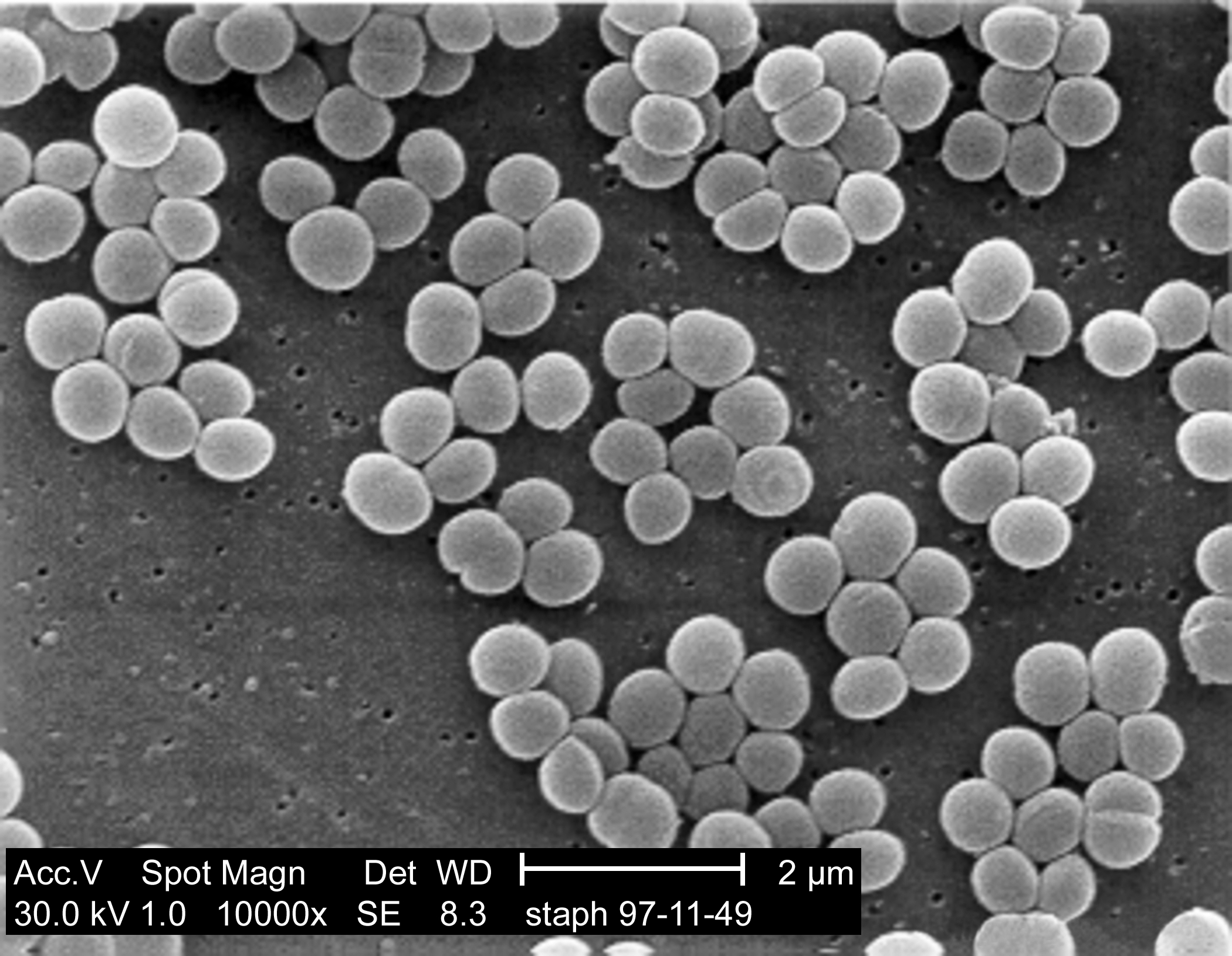
This is the strategy used by Methicillin, an antibiotic that used to be talked about a lot a while ago (it's the 'M' in MRSA) but has been neglected by the media lately in favour of swine-flu and other viruses. Methicillin is a B-lactam antibiotic, which means that it binds to one of the enzymes involved in cell wall metabolism, blocking its active site. More specifically, it binds to the enzyme that creates the cross-links between the glycopeptides (PBP2). No new cell wall can be created, and therefore no more bacteria.

Resistance to this takes several forms. MRSA simply uses a variant of the enzyme, with a deeper active site, so that while the cell-wall precursor substrate can bind, the antibiotic cannot. Protection from a wide variety of different B-lactams can be achieved by B-lactamases, bacterial enzymes which break down the antibiotics. Multi-efflux pumps also exist, these are proteins that span the bacterial cell wall and essentially pump out any antibiotics that make their way into the cell before they can cause any harm.

Vancomycin is the drug that is still most commonly used against MRSA, although some resistance is (as always) beginning to arise. Unlike methicillin, vancomycin does not bind to any bacterial enzymes, instead it binds directly to the cell wall precursors. The part it binds to is shown below, as a close up from the earlier diagram of the cell wall:The incredibly inexpertly added D-ala in red at the bottom shows the precursor form of this section of the cell wall (Ala, Glu and Lys are the short-hand form of amino-acids, so this is just a short protein chain. The L- D- labels show what form the amino-acids are in). The vancomycin binds to the final D-ala-D-ala, preventing it from being processed and halting construction of the cell wall.

Two different methods of preventing cell wall growth; one antibiotic binding to the enzyme, the other to the substrate. And both, sadly, have been defeated by resistance already. In the case of the B-lactams, a wide variety of resistance mechanisms exist, from actively destroying the antibiotic, to pumping it out the cell, or bypassing the enzyme completely. In the case of vancomycin, some bacteria have started producing peptide chains ending in D-ala-D-ser, or D-ala-D-lac, and there have been reports of VRSA from America.

The pathway for creating bacterial cell walls contains multiple steps, and both methicillin and vancomycin halt just one of these, the cross-linking of the short peptide chains near the end. Bacitracin, another cell-wall directed antibiotic, works further upstream in the pathway. I've just been given a whole stack of papers on bacitracin by my supervisor...so I'll probably be writing more about that in the future!

Bacteria that use antibiotics...for food!

ResearchBlogging.orgAntibiotic resistance is by now a well-known phenomenon. Resistance is carried in both antibiotic producing bacteria to protect themselves from their own weaponry, and the soil bacteria they attack, in an attempt to defend themselves. The sudden influx of pharmaceutical antibiotics has encouraged the spread of resistance to human pathogenic strains, leading to the so-called 'superbugs' seen in the media such as MRSA and vancomycin-resistant C. difficile.

However researchers at Harvard found that not only are some bacteria able to neutralise the threat of antibiotic resistance, they actually use antibiotics as a food source. Not only that, but they were capable of using antibiotics as the sole carbon source. The table below (taken from the reference at the end of the post) shows the survival of bacteria on antibiotics using samples from three different types of soil, Farmland (F), Urban (U) and Pristine (P - soil from non urban areas with minimal human contact for 100 years):

The antibiotics used include natural, synthetic and semi-synthetic molecules, all all of which could be used by bacterial species as a carbon source. Even more interestingly (or alarmingly) the antibiotics were at concentrations of 1g/litre, 50 times higher than the concentration normally used to test for resistance.

The 'pristine' soil is the one that the researchers found the most interesting, as the general expectation was that this area would contain fewer antibiotic-eating bacteria, having had minimal interaction with people and pharmaceutical antibiotics. However the data showed no noticeable difference, despite not being in contact with human-designed antibiotics, the bacteria are meeting plenty of bacterial-based antibiotics, and adapting to use them for food.

The big question of course is Will it Spread? Around the quarters of the isolated strains belonged to orders containing clinically relevant strains such as Salmonella and E. coli, meaning that hypothetically at least antibiotic consumption should be able to spread. On the other hand, actual consumption of antibiotics is unlikely to provide a greater evolutionary advantage than just resistance, and will confer a larger metabolic load on the bacteria. Although the pathways of antibiotic metabolism have not yet been fully determined, the first few steps seem to be similar to well-known resistance mechanisms (particularly in penicillin consumption). One conclusion, therefore, is that only part of the metabolic pathway would be (or already has been) passed on to pathogenic organisms, enough to provide resistance without placing unnecessary metabolic burdens on the cell.

Hat tip to Byte Size Biology for alerting me to the paper.

--

Dantas, G., Sommer, M., Oluwasegun, R., & Church, G. (2008). Bacteria Subsisting on Antibiotics Science, 320 (5872), 100-103 DOI: 10.1126/science.1155157

Cell wall under attack - bacterial response to antibiotics

ResearchBlogging.orgI took a quick break away from synthetic biology and DNA synthesis research the other day, to dive back into my happy little world of antibiotic research, in preparation for my new project in October. I'll be working with Streptomyces bacteria again, which after a whole summer of E. coli I'm quite looking forward to. What I'll be doing with them is examining the response of the cell wall to antibiotics.

The bacterial cell wall is made up of glycopeptide molecules (sugars and proteins joined together) and surrounds the whole cell. Without it, bacteria swiftly loose their integrity and salt-balance across the membrane, which is why many antibiotics target the cell wall in order to kill bacteria. Both for antibiotic resistance, and for surviving conditions that could damage the cell wall, bacteria have a system of monitoring the state of the cell membrane and responding quickly to any changes.

The system that was discovered in Streptomyces coelicolor (which I'll be working on) was named the sigE system, and consisted of an operon (string of genes) encoding four genes:

SigE encodes for a sigma-factor, a protein used in bacteria to switch on certain sets of genes. The cseA codes for a cell membrane lipoprotein, possibly used in a sensor system, while cseB and C are a two-component signalling system (very common in bacteria). CseC is a sensor (a histadine protein-kinase sensor for those who are interested) while cseB is the response regulator, acting out a response when it receives a signal from cseC.

And now...the science :)

In order to test that this operon was involved in cell membrane responses to antibiotics the lab carried out a variety of experiments, all producing evidence that lead towards this conclusion. The main experiments were as follows:
  1. Removing the sigE operon and placing it on a separate plasmid, that activated resistance to Kanamycin. The bacteria were then plated on agar containing antibiotics and challenged with a kanamycin disk. Cell wall attacking antibiotics induced kanamycin, whereas antibiotics that attacked (say) the ribosome didn't.
  2. Keeping the sigE in its original chromosomal context, the group then challenged it with different concentrations of vancomycin (an antibiotic which attacks bacterial cell walls). They then measured the level of the sigE operon proteins being produced in the cell. Higher concentrations of vancomycin, lead to more proteins.
  3. Going back to the sigE-kanamycin resistant protein, they tried knocking out the sigE promoter, effectively switching all these genes off. The effect seen previously disappeared.
  4. Leaving the lab, they then did some computational work, scanning the database to see what genes the sigE sigma-factor actually switched on. They found a group of 12 genes, all of which coded for cell-wall synthesis enzymes.
All of this leads up to some pretty conclusive evidence - in case of cell wall damage, the sigE operon is switched on. The interesting thing is, is that this isn't just a response to antibiotics either. It is highly unlikely that the system is able to respond to every different cell-wall destroying antibiotic, instead, the response is triggered by cell-wall intermediates, or degradation products that signal "Help - cell wall is being destroyed!" and switch on the sigE response, which produces proteins to mend it again.

But there are still a lot of unanswered questions. What is the cseC actually sensing? What is the exact purpose of the cseA? Why produce both a sigma-factor and a heafty response pathway? Which intermediates are used for activating? And, most importantly, can we hijack this somehow to kill bacteria?

I can't wait to get to work with it :D

---

Hutchings, M., Hong, H., Leibovitz, E., Sutcliffe, I., & Buttner, M. (2006). The E Cell Envelope Stress Response of Streptomyces coelicolor Is Influenced by a Novel Lipoprotein, CseA Journal of Bacteriology, 188 (20), 7222-7229 DOI: 10.1128/JB.00818-06

Hong, H., Paget, M., & Buttner, M. (2002). A signal transduction system in Streptomyces coelicolor that activates the expression of a putative cell wall glycan operon in response to vancomycin and other cell wall-specific antibiotics Molecular Microbiology, 44 (5), 1199-1211 DOI: 10.1046/j.1365-2958.2002.02960.x

Jacobs C, Frère JM, & Normark S (1997). Cytosolic intermediates for cell wall biosynthesis and degradation control inducible beta-lactam resistance in gram-negative bacteria. Cell, 88 (6), 823-32 PMID: 9118225