Field of Science

Showing posts with label bacteria. Show all posts

Bacterial Phylomon!

A while ago a conservationist called Andrew Balmford wrote a letter in Science pointing out an intriguing fact that many parents are already well aware of. Namely that while many young children are able to name and recognise over 100 species of Pokemon (fake creatures from a magical land used for a card game and now a good number of computer games) they are usually woefully ignorant of the types of birds that fly around their houses.

The Science Creative Quarterly then got the idea that if children like playing with cards of wierd and wonderful creatures why not make actual cards of real-life creatures for them to play with? Thus the phylomon project was born, and now boasts an impressive collection of cards showing diverse creatures from all over the planet.

Will it work? Well quite a few parental and teacher hopefuls have downloaded the cards, so presumably they're getting something out of it. There are rules for games, and some kids have made their own cards, but I'm not sure it will ever have quite the same appeal as pokemon. Or even dinosaurs. Dinosaurs were the original "why is my kid able to remember a hundred difficult names but can't remember what her sister's called?" item. Their fascination is that they are powerful, mysterious and almost magical. Pokemon even more so. Face it - a tree is just not as exciting as a velociraptor.

Another thing that I find harder to relate to is that while the cards do have beautiful illustrations the animals have no humanly identifiable expressions. For me, the main draw of Pokemon was that every one of the things had both a name, a load of sweet attacks, and an actual personality. The cards had expressions. Charmeleon was a sulky teanager, Bulbasaur was an overly-large slightly shy kid, Rattata was fierce and always on edge for a fight. Maybe it's just me, but I loved the fact that they each had a history, and a story and a set of almost human feelings and emotions.

So in the spirit of joining in, I have made some bacteria cards. And in the spirit of Pokemon, they all have eyes, expressions, and attacks...


They probably would have been better if I had any drawing ability. Or more than three pens.

Don't miss my first SciAm Blog post about bacteria that communicate with electricity!Link

Plant Defence 3 - Acquired Resistance

ResearchBlogging.orgIn posts one and two of this mini-series I explored how plants can defend against bacteria by releasing dangerous chemicals and by killing off cells. This post looks at how surviving one bacterial attack can make plants more able to survive subsequent ones with both local and systemic acquired resistance.

Locally acquired resistance is the simplest to manage, and provides a clear advantage. If cells have been attacked once it makes sense to defend them in case of a second attack. Plants achieve this by strengthening the cell walls in cells that have survived the bacterial attack. Experiments adding elicitors (bits of bacteria that stimulate the plant pathogen receptors) to plant cells showed that proteins in the cell wall became oxidatively cross-linked as they sense the bacteria. Interestingly the molecule responsible for this is hydrogen peroxide, one of the molecules also involved in the cell-death response discussed in post two. If it doesn't kill the plant, it makes it stronger.

The cell membrane is the blue box at the bottom, whereas the cell wall is the light blue rods in the middle. It is the cell wall which is strengthened. Image from wikimedia commons.

This response is all very well for plant cells which happened to be near the site of infection, but what about the rest of the plant? Is it possible for cells on the other side of the plant to be warned and ready for a pathogen attack? Despite the inability of plant cells to move, the answer surprisingly is yes. Cells at the site of infection can release a chemical called salicylic acid which moves through the plants vascular system (the system which also delivers sugars and other important nutrients to all parts of the plant).


The chemical structure of salicylic acid, which is chemically similar to the active component of aspirin.

Following an infection, the levels of salicylic acid were found to rise dramatically in cells around the zone of infection, before spreading through the rest of the plant. This isn't a species specific response either but one found in many different species; grafting parts of one plant onto another did not stop either plant from acquiring resistance. In response to the salicylic acid signal cells start accumulating small amounts of hydrogen peroxide, which can lead to the same cell wall strengthening seen around the area of infection.

As well as salicylic acid it has also been suggested that infected areas of the plant can release the volatile molecule methyl salicylate, commercially known as oil of wintergreen. Rather than travelling through the plant this signal is airborne, allowing transmission not just to other parts of the plant, but to neighbouring (and therefore likely to be related) plants as well. As the only difference between these two signalling molecules is the addition of a small CH3 group, the methyl salicylate can easily be converted back into salicylic acid once it reaches the cells where it can cause the same downstream response.

If anyone was wondering quite why I've suddenly been into plants part of the reason is that the BBC is showing a program called "Botany - a blooming history" and I've been catching the episodes. Despite the slight naffness of the title, it's actually a really good program showcasing experiments, personalities, and the scientific method as it unfolds the history of plant science. You can catch the episodes here on iPlayer.

---
Brisson, L., Tenhaken, R., & Lamb, C. (1994). Function of Oxidative Cross-Linking of Cell Wall Structural Proteins in Plant Disease Resistance The Plant Cell, 6 (12) DOI: 10.2307/3869902

Durrant, W., & Dong, X. (2004). SYSTEMIC ACQUIRED RESISTANCE Annual Review of Phytopathology, 42 (1), 185-209 DOI: 10.1146/annurev.phyto.42.040803.140421

Shulaev, V., Silverman, P., & Raskin, I. (1997). Airborne signalling by methyl salicylate in plant pathogen resistance Nature, 385 (6618), 718-721 DOI: 10.1038/385718a0
---

Follow me on Twitter!

Plant defence 2 - Honourable Suicide

ResearchBlogging.orgThe first post of this mini-series covered how plants can defend themselves against bacterial attack by releasing chemicals, either on a regular basis or as a specific response to the attack. This post will explore the hypersensitive response, which allows plants to rapidly kill of cells around the area of infection, starving the bacteria of nutrients to prevent it spreading. The end result is a small area of dead plant matter, with the rest of the organism unaffected.

One of the main differences between plants and animals that I flagged up in the last post is that plant cells don't move. The use of the hypersensitive response shows another; plants have a very non-determinant structure. Animals will grow towards a clear well defined shape and once they get it, they stick with it. Your body does change as you grow older, but it's not about to grow an extra leg. Plants on the other hand may have determinant structures within them, such as leaves or flowers, but the overall organism can just keep growing for as long as it needs to. If a leaf is lost through disease, the plant can just grow a new one, or several new ones.

These leaves are expendable. Your arm is not. Image from wikimedia commons.

Because of its non-determinant nature it is a lot easier for the plant to kill parts of itself off in order to stop an infection spreading. One way that the hypersensitive response does this is by the production of large numbers of reactive oxygen species in cells surrounding the site of infection. These include hydrogen peroxide, and various hydroxide and oxygen containing free radicals. Free radicals are species with one unpaired electron and therefore are extremely reactive and extremely dangerous. These free radicals lead to chain reactions that can break down lipids in the membrane, inactivate enzymes and generally roll around like a loose canon causing havoc within the cell.

As well as reactive oxygen species, the cell also experiences large ion fluxes, as potassium and hydroxide ions flood into the cell and hydrogen and calcium ions flood out. These result in the cell releasing any stored toxic compounds it might have (which may also help to kill the bacteria) and may serve to integrate the mitochondria into the process of cell death (see reference one). As mitochondria are crucial in coordinating the programmed cell death of animal cells it would be surprising if they did not play some part in the controlled destruction of plant cells. The actual sequence of destruction varies from plant to plant, but the overall result is the same, and area of dead plant tissue within the still healthy surviving plant.

A leaf infected with tobacco mosaic virus, showing lighter areas of dead leaf interspersed with the green areas of normal growth. Image from wikimedia commons.

The plant hypersensitivity response can (if you want it to) be considered analogous to the human innate immune response, in that it occurs directly in response to a bacterial attack, and it occurs only at the site of bacterial infection. Plants, however, also have ways of making more long-term changes to protect against bacterial attacks in the future both at the site of the old reaction and throughout the whole plant. How the plant achieves this, without any cellular movement, will be the topic of the final post in this mini-series.

---
Lam, E., Kato, N., & Lawton, M. (2001). Programmed cell death, mitochondria and the plant hypersensitive response Nature, 411 (6839), 848-853 DOI: 10.1038/35081184

Pontier, D., Balagué, C., & Roby, D. (1998). The hypersensitive response. A programmed cell death associated with plant resistance Comptes Rendus de l'Académie des Sciences - Series III - Sciences de la Vie, 321 (9), 721-734 DOI: 10.1016/S0764-4469(98)80013-9

Taiz, Zeiger, Plant Physiology, third edition Sinauer Associates 2002.
---

Follow me on Twitter!

E. coli the enemy

It's been in the news a lot, but for those who've missed it there's a particularly deadly strain of E. coli that has reared its head in Germany. The source of the bacteria is not yet known although both cucumbers and beansprouts have been blamed, and German authorities are advising people to stay away from raw leafy vegetables. Vegetables don't usually harbour their own strains of E. coli, but the thought is that animal manure (which most definitely does contain E. coli) used to fertilise the vegetables may be carrying the deadly strain.

This plate is more deadly than you can possibly imagine

I talk about E. coli a lot in my blog as I use it for most of my experiments. The strain I use is called K12 and is completely harmless to humans. It's also used in sterile conditions and not allowed to leave the lab. The strain that's causing panic in Germany is called O104:H4 which is an enterohemorrhagic strain which can cause bloody diarrhoea and also attack the kidneys. Working from news sources (as there don't seem to be any papers out yet!) it seems to have picked up the DNA with a kidney damaging toxin as well as having proteins that help it to stick strongly to the intestinal cell wall. Other bloggers have looked at this in more exact detail (and shared their findings, which is pretty awesome), including which parts of the DNA have changed in this new strain.

The labelling isn't just arbitrary either, it tells you exactly what antigens the bacteria is carrying, and antigens are the things that the immune system recognises to help your body attack the disease. There are three different types of antigens in the E. coli species; O (in the new deadly strain), H and K (in my lab strain). The letter and number after the : mark "O104:H4" refers to the flagella antigen. Strains which share the same antigen types will have similar patterns of virulence.

As well as trying to understand the genetic code and how this strain differs from other deadly and non-deadly E. coli, scientists are also trying to find exactly where the bacteria are coming from, testing vegetable sources in an attempt to isolate it. Both cucumbers and beansprouts have now been tested for the strain isolated from patients, and both have shown negative results.
Understanding where the deadly strain arises from is less useful in terms of the scientific investigation, but crucial for stopping the disease from spreading, and helping to understand how to avoid such strains developing in the future.

If you're worried about the vegetables you're eating then the best advice I can give is to cook before eating. E. coli are not able to withstand the high temperatures of cooking and unlike bacteria such as clostridium they do not release toxins onto the food. Clostidium produce exotoxins directly onto your food whereas E. coli produce endotoxins once they get into the body. For the more sciencey minded, it's Gram positive bacteria which tend to produce exotoxins, whereas Gram negatives produce the endotoxins.

E. coli image from Wikimedia commons

It's only once the E. coli enter your body that they start producing 'toxins' and these toxins are usually parts of the bacteria as they are broken down by the immune system. If you manage to destroy the bacteria before eating the food, then it will be pretty much safe to consume. Note that this technique does not work for exotoxins, which are actively secreted by the bacteria (Gram positives like secreting things) onto food before you eat it.

Studying bacteria does sometimes feel like a little niche set slightly apart from the real world, which is full of lumbering eukaryotes making complex non-rational interactions. News like this an interesting reminded that bacteria don't just affect health, but also have strong economic and political implications. Spanish cucumber salesmen are trying to sue Germany, Russia's getting all smug about EU health regulations, and German tourism is being affected. All because of E. coli.

Social Evolution in Bacteria - SGM series

This is the fourth post in my latest SGM series.

ResearchBlogging.org
The social behaviour of bacteria is something that I get very excited about. From the wolf-pack hunting strategies of Myxococcus xanthus to the terminal differentiation of cyanobacteria, it's something that I never get tired of writing about. As well as providing interesting quirks of bacterial behaviour, living within a colony also gives new scope for exploring the evolution of bacteria; not just as single entities but as a fully functioning social group.

One of the differences of living within a social colony as opposed to alone means that altruistic-type behaviour has to be adopted. Bacteria living within a biofilm need to excrete the sticky goo that holds the biofilm together, which is problematic because synthesising and secreting goo takes up a lot of energy. So within this colony, there will be 'cheaters' - those bacteria that live in the surrounding goo produced by others, while making none themselves.

A bacterial biofilm, showing individual bacteria in green. Image taken from the FEI website, shown there courtesy of Paul Gunning, Smith & Nephew

As with all colonies, cheating might benefit the individual but has no benefit for the colony as a whole. Too many cheaters and there won't be any biofilm. And recently an even more subtle form of cheating has been shown within the biofilms of the bacteria Pseudomonas aeruginosa, with bacteria that don't just refuse to make vital sticky chemicals, but also abstain from the entire process of forming a biofilm.

Bacteria use a complex communication system called quorum sensing in order to determine how many other bacteria they are surrounded by. Once enough bacteria are present, all signalling their existence, the biofilm will start to form. However some bacteria isolated from the biofilm were shown not to be taking part in any quorum sensing at all. Quorum sensing appears to be quite a burden for a growing cell - cells with the quorum sensing genes knocked out tend to grow a lot faster that the socially conscious cells that allow biofilms to form.

The paper that goes through this (reference one) highlights it as a form of social cheating, with bacteria avoiding quorum sensing to benefit themselves while mooching off the quorum sensing behaviour of others. I'm not entirely certain that this is the case though. It may just be an good example of job allocation within the bacterial society. Clearly not all bacteria are required to be continually quorum sensing, so why should they all have to? Would it not be more sensible to have some exempt from that task, so that they can concentrate on growing, dividing, and spreading the colony? This may be more a case of tax-breaks than of benefit-cheats.

Social evolution doesn't just take place within species, but also between them, and like every other organism bacteria are in a constant state of coevolution with both their 'prey' and their predators. Most predator-prey interactions take long periods of time to study, but the beauty of bacteria is that you can go through several generations in the course of one week's growth. Studies of the bacteria Pseudomonas fluorescens and its bacteriophage parasite showed that both the bacteria and the bacteriophage evolved far quicker when interacting together than they did when competing against a non-changing opponent.


Bacteriophage surrounding a bacteria. Image from wikimedia commons

'Evolve' here means that the bacteria and the bacteriophage showed a greater change in their genetic makeup, and a greater genetic divergence from bacteria not pitted against the phages. Unsurprisingly, the genes that changed the most were those involved in host-phage interaction. This study (reference 2) is also a great example of the usefulness of whole genome sequencing. Whole populations of bacteria and phage were allowed to evolve both together and separately and then just sent away for sequencing with the results analysed at the end.

You really can't be an anti-evolutionist while studying bacteria. They just do it so damn quickly and often you can see it happening.

---
Sandoz, K., Mitzimberg, S., & Schuster, M. (2007). From the Cover: Social cheating in Pseudomonas aeruginosa quorum sensing Proceedings of the National Academy of Sciences, 104 (40), 15876-15881 DOI: 10.1073/pnas.0705653104

Paterson S, Vogwill T, Buckling A, Benmayor R, Spiers AJ, Thomson NR, Quail M, Smith F, Walker D, Libberton B, Fenton A, Hall N, & Brockhurst MA (2010). Antagonistic coevolution accelerates molecular evolution. Nature, 464 (7286), 275-8 PMID: 20182425
---

Follow me on Twitter!

Life at zero growth rate - SGM series

This is the third post in my latest SGM series.

ResearchBlogging.org
One of the first topics that I learnt in Biology was that there are two types of things; living things, and dead things. Living things are given a whole host of distinguishing characteristics (growth, reproduction and, my favourite, irritability) where as dead things are defined as everything else. Biology was usually defined as the study of living things.

As I grew older, I found that there were many complications to this neat little classification. Viruses - which are neither fully living, nor properly dead. A whole organism can be dead, despite the fact that many of its cells are still alive (how alive is a freshly killed animal? Or the flowers in a vase?). And of course what is for me the most intriguing case, that of dormant bacteria.

Dormancy is an odd state to be in. A dormant organism shows none of the signs of being alive. It does not eat, grow or divide (although some very basic metabolic processes may still continue). It shows no response to any outside stimulus, and can often be placed in conditions that would
lead the living organism to perish, such as extremes of temperature and pressure. Yet somehow just one simple stimulus can cause this previously dead looking organism to spring magically back into life.

Bacteria are not the only things that can go dormant. Some
animals can as well, the most famous example being tardigrades -the thing shown on the right that looks a bit like a plushie made by Tim Burton (image from wikimedia commons). Yeast are well-known for forming dormant spores, and it can be argued that a seed is technically a dormant plant, just waiting for water to be added to bring it back to life.

One of the most medically important dormant bacteria is Mycobacterium tuberculosis which infects humans and leads to TB. One of the reasons for its pathogenicity is that they can go dormant, both outside the body (which makes them hard to shift from a hospital) and inside the body, after the primary infection (which makes them even harder to shift from inside a human body).

Although the latent cells can remain within the body for many years, sometimes never coming back from dormancy at all, ideally there should be some signal to bring them back to life. These signals are known as "resuscitation-promoting factors" or RFPs. These RFPs are required for virulence, and to bring the bacteria back from dormancy, but are not necessary for the growth and proliferation of cultures in the lab.

Within human tissues, and throughout the cycle of the disease, you can track these RFPs to try and get an insight into what the bacteria is up too, and when it may move from latent periods to periods of active growth. As well as being useful for tracking the course of infection, this might also have therapeutic implications. If you can convince the bacteria not to come out of dormancy then you have an infection state that might not be completely curable but is at least controllable.

How organisms survive in a state of dormancy, and indeed how they ever come out of it, is a subject I find really fascinating. I'm unlikely to ever get to do much research on it (because as fascinating as it might be screwing around with my little bugs till they do what I want is endlessly more fun) but I'll probably have a good few more posts writing about it and exploring how it works.

---
Kana BD, Gordhan BG, Downing KJ, Sung N, Vostroktunova G, Machowski EE, Tsenova L, Young M, Kaprelyants A, Kaplan G, & Mizrahi V (2008). The resuscitation-promoting factors of Mycobacterium tuberculosis are required for virulence and resuscitation from dormancy but are collectively dispensable for growth in vitro. Molecular microbiology, 67 (3), 672-84 PMID: 18186793

Davies AP, Dhillon AP, Young M, Henderson B, McHugh TD, & Gillespie SH (2008). Resuscitation-promoting factors are expressed in Mycobacterium tuberculosis-infected human tissue. Tuberculosis (Edinburgh, Scotland), 88 (5), 462-8 PMID: 18440866

Guarding Microbial diversity - SGM series

This is the first of the Spring 2011 SGM series; where I steal random topics from the Society for General Microbiology Spring Conference and write about them in my blog. It should be noted that I am in no way affiliated to the Society, I'm just currently not rich or scientific enough to go to their conferences. (next year...)

I was quite intrigued when I saw this topic, as microbial diversity has always seemed to me to be a little bit like rats. Interesting to the people that study them, irritating and potentially hazardous to those who don't, but not really in need of any special protection. Bacteria evolve quickly, and share DNA easily, forming many, many diverse species capable of occupying a wide variety of niches.

However while bacteria are indeed very diverse and happy to remain so, the challenge comes in cataloguing all of that diversity. New bacteria need to be examined, named, placed in a taxonomic group, and preferably stored so that if anyone has a particular urge to work on (say) a specific type of hydrogen-eating swamp bacteria, they can find a sample of it and do that work.

What I hadn't realised was that there are quite a few places that are designed to store bacterial cultures. One such place is the DSMZ collection in Germany which boasts over 20,000 cultures of assorted microorganisms. These are stored as dried samples, not alive yet easily able to resuscitate. For a small fee, you can order a sample from them, together with instructions as to how to bring it back to life and culture it within a laboratory. Without storage facilities such as these, it's easy to see how interesting new bacteria would simply get lost, due to freezer melt-downs in labs, or people discarding the wrong samples.

It's not just the finding and the storing of bacteria that are vital in order to maintain scientific knowledge of their diversity, you've also got to name the things. After all, an unlabelled catalogue is no use at all. And with the name comes characterisation - a whole list of the properties and behaviour of the bacteria down to as much biochemical information as is feasible. The speed and accuracy of full-genome sequencing does make this a lot easier, but there are still many properties that depend on more than just the genes. A bacterium might possess the gene for (say) iron metabolism, but that doesn't mean it uses it all the time, or indeed at all. Knowing the genome sequence also makes it a lot easier to quickly place a bacterium into an existing group or species. Although bacteria do share DNA between each other, recently acquired DNA can usually be distinguished from the core genes that mark the species.

UK strains can be acquired through the Health Protection Agency, which is an aggregation of four previously separate culture collections. The bacterial arm of it has around 5000 different bacterial cultures. I did do a quick check as to whether I could get a small sample of S. erythraea using my Debit Card, but you need to be officially registered before they start handing out the bacteria!

It's quite strange to think that these talks are actually taking place up in the north of the country while I'm writing this. Next year though, I'm aiming not just to turn up, but actually bring a poster with me, to show off some of my own work.

New SGM series!

The Society for General Microbiology runs regular conferences that concentrate exclusively on the world of the very small. Although I am currently not a member :( I have been in the past, and plan to be again next year!

The conferences are always very good, and last august I bemoaned not being able to make one. This spring I am also not able to attend as I've only just come back from my honeymoon, and in the general scheme of work-life balance it would be a very sad and hardcore worker who would put an actual wedding second to marriage.

The last SGM conference that I missed, I decided to do a nerdy little one-women conference of my own, here at Lab Rat. And it worked so well that this time I'm going to try and do it again. One post every two days, covering the following topics in no particular order:

Seeing the cell through the “eyes” of the virus
Guarding microbial diversity
Vaccines
Insect symbiosis
Life at Zero Growth Rate
Maths & microbes - heh that'll be fun!
Social evolution in micro-organisms

Seven topics, which should take me two weeks to get through. It'll be a good way to get my self back into blogging and back into the exciting world of bacteria. It should be an interesting fortnight!

Signals for Infection

ResearchBlogging.orgNeisseria meningitidis is a bacteria which lives in the throats of around 30% of the human population. In most cases it causes no problems at all and just exists as a normal part of the throat microbial flora. In some patients however it can start to colonise the bloodstream and brain, leading to cases of septicemia and meningitis which are highly dangerous and can be fatal.

The invasion starts with individual bacteria, which adhere to the epithelial cells that cover the inside of the throat. They then start to divide and proliferate to form large aggregated colonies. Within these colonies they are connected to each other, and to the epithelial cells, by protrusions from the bacterial cell surface called pili which are shown below for a wild-type (i.e un-genetically modified) Neisseria meningitidis:
Image taken from the reference below. The arrow points to one of the pili, and the insert shows a close-up of it.

These pili are often modified by the attachment of small molecules to the pili proteins, including the molecule phosphoglycerol (shown on the right for those interested in structure). To test the effects of the addition of phosphoglycerol, the researchers found which gene caused the addition of this molecule onto the pili (the pptB gene), and removed it from the cell. Without the pptB gene there was still the same number of pili around the cell, but they were not clumping together as much. Instead of the thick fibres seen in the wild type above (caused by large bundles of pili) only little stringy fibres were seen. These thin spindly fibres show that without the addition of phosphoglycerol, the pili cannot clump together.

This is important medically as Type IV pili bundle formation and N. meningitidis aggregation for infection are linked. Interestingly it was not the aggregation that was affected by removing the phosphoglyerol but the ability of individual bacteria to leave the aggregate to infect other parts of the body. In wild-type bacteria, the pptB gene is strongly activated only after several rounds of division within the aggregate, so it looks like the addition of phosphoglycerol acts as a switch, communicating to the bacteria that enough of them have aggregated and it is now time to leave. If the pptB is activated due to large numbers of bacteria it could act as a communication of the population density - signalling to the individual bacteria that the current location is far too crowded, and it has better chances of survival if it leaves.

---
Chamot-Rooke J, Mikaty G, Malosse C, Soyer M, Dumont A, Gault J, Imhaus AF, Martin P, Trellet M, Clary G, Chafey P, Camoin L, Nilges M, Nassif X, & Duménil G (2011). Posttranslational modification of pili upon cell contact triggers N. meningitidis dissemination. Science (New York, N.Y.), 331 (6018), 778-82 PMID: 21311024
---
Follow me on Twitter!

Coloured bacteria vs. Angry birds

For those who didn't catch the news when it first came out, the iGEM project I carried out in the summer of '09 has been nominated for a Design Award! The Brit Insurance Design of the Year 2011 to be precise.

I'm very excited about it, even though I don't really feel like a designer (I don't own a singlething made by Apple). We're up against some pretty intense competition, such as the Angry Birds phone game, Rock band 3 and quite a few phone apps. Still, it's pretty awesome to be nominated, and I'm looking forward to seeing all the rest of the project displays.

Our two wonderful designer-friends James King and Daisy Ginsberg have put together a great video explaining the project. It's got some great animation, and clips from a radio interview with one of my fellow lab rats, and even some pictures of the infamous Orange Liberation Front...

E. chromi from Alexandra Daisy Ginsberg on Vimeo.

Targeting dormant bacteria

ResearchBlogging.orgAntibiotics are effective against bacteria because they target and knock out specific functions that are vital for bacterial survival. As most bacterial infections involve rapid growth and division of the invading bacteria, many commercial antibiotics currently target metabolically active cells, by blocking enzymes needed for growth, reproduction, or cell wall synthesis. While these will kill acute bacterial infections they are often far less effective against dormant bacteria in longer-term persistent infections.

Rather than targeting metabolic enzymes, the current strategies being explored to combat dormant bacteria target either the membrane, or membrane bound proteins. Both of these approaches destabilise the bacterial membrane and help to break the cell apart and can act against processes such as energy synthesis which occur in both active and dormant cells.

a=targeting important metabolic proteins in the membrane. b=targeting the actual cell-membrane. Picture is copywrite me :p

In eukaryotic cells, such as the cells of plants and animals, the enzymes that create energy for the cell are kept safely hidden away in specialised intracellular compartments, such as mitochondria. As energy production requires an ion gradient across a membrane, these compartments all have sets of internal membranes. Bacteria however do not have this luxury, and instead have all their metabolic enzymes in the outer cell membrane, as this is the only membrane they have. Inhibitors of energy metabolism can therefore bind directly to target enzymes in the membrane involved in the production of energy. This can be highly effective against cells whose interior is hard to get into, such as Mycobacterium tuberculosis which lurks inside tuberculosis granulomas. Even in the absence of growth, cells still require a minimal energy input to survive, so blocking off these enzymes kills both dormant and active cells.

Drug developed to help combat TB by attacking cell membrane metabolic enzymes. This drug is currently in stage three clinical trials.

The membrane-targeting drugs act directly on the lipid bilayer that surrounds the bacterial cell, breaking it up and destroying the bacterial cellular integrity. Although human cells are also surrounded by lipid bilayers they have fewer negatively charged phosopholipids and also contain cholesterol (not present in bacterial membranes) allowing membrane-targeted drugs to be specific for human pathogens rather than killing surrounding human cells. The drugs that are used to attack the cell wall can vary hugely in size and structure but they all share one common property; they are highly lipophilic (i.e they are attracted to lipids). This allows them to interact with the cell membrane and break it apart.
Lipophilic drug capible of targeting bacterial cell membranes

There’s something about those molecular diagrams of drugs that I love. I think it’s my biochemical background. I’m never totally happy with a schematic until I can see how the chemicals are interacting on a molecular scale.

As well as being useful against dormant bacteria these new antimicrobials show promise as strategies for dealing with arising antibiotic resistance. Bacteria can evolve to cope with as many challenges as are thrown at them, but hopefully it should take them a little longer learn to survive entirely without a cell wall…

Although there are some that can do that already.

---
Hurdle JG, O'Neill AJ, Chopra I, & Lee RE (2011). Targeting bacterial membrane function: an underexploited mechanism for treating persistent infections. Nature reviews. Microbiology, 9 (1), 62-75 PMID: 21164535

---
Follow me on Twitter!

Guest post - Microbes and Madness

This guest post comes from my fiancé who is a Psychiatrist. I've been very excited about this post for a while, because unlike me, he is a published author who has written a book on Consciousness and the philosophy of mind.

Microbes and Madness

At first glance, it would be reasonable to assume that my profession and that of the author of this fabulous blog are poles apart. However, everything in nature has a connection, and so it is not surprising to discover a fascinating area where psychiatry and bacteriology overlap.

A broad range of pathogens are known to cause psychiatric sequelae, including worms (neurocysticercosis), protozoa (cerebral malaria, toxoplasmosis), viruses (HIV, herpes simplex encephalitis, rabies), prions (Creutzfeldt-Jakob disease, kuru), and, of course, bacteria (neurosyphilis, Lyme disease, post-streptococcal syndromes). However, in the spirit of this blog, this post will be focusing on bacteria.

There are essentially four mechanisms through which bacteria cause psychiatric symptoms in humans:

I. Bacteria can infect the central nervous system and cause direct damage to brain cells.
II. Bacteria can trigger a powerful systemic inflammatory response that results in a disruption in brain function.
III. Bacteria can trigger an adaptive immune response which produces antibodies that cross-react with host central nervous system proteins.
IV. Bacteria can be the objects of a phobia.

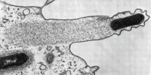
Syphilis and Lyme disease are examples of infections which involve the first mechanism. Syphilis is caused by spirochaetes of the species Treponema pallidum, and is sexually-transmitted. Lyme disease is caused by spirochaetes of the genus Borrelia, and is vector-borne, with ticks from the genus Ixodes being the commonest vector. Both diseases are associated with widespread dissemination of infection of multiple organ systems, and are notorious for their protean manifestations.

The range of possible psychiatric presentations is vast. Syphilis, in particular, can mimic any psychiatric syndrome, and was a common diagnosis in psychiatric inpatients a century ago. The possible range of presentations include delirium, dementia, psychosis, mania, and personality changes. Lesions of the frontal lobes are associated with personality changes and disinhibited behaviour, whereas those of the temporal and parietal lobes are associated with cognitive decline. Lyme disease can also mimic several different psychiatric syndromes, but typically affects the limbic system, causing disorders of emotional regulation, including panic attacks, phobias, depression, and obsessive-compulsive behaviour.

The second mechanism listed refers to sepsis-associated delirium. No human organ system is a closed system, including the central nervous system. Bacterial infections with a focus outside the outside the brain are capable of causing a systemic reaction, which affects the brain. The result is an acute confusional state, or delirium.

Common causes are pneumonias and urinary tract infections, although infections of other organ systems are also frequently implicated. Delirium presents as a transient global disorder of cognition. Typically, there is clouding of awareness, disorientation, impaired attention, fluctuating alertness with agitation or drowsiness, hallucinations, illusion, and vague delusions. The state is thought to be caused by a global disruption of brain function, which may result from the effects of a systemic inflammatory response to infection. These effects may include systemic vasodilation causing cerebral hypoperfusion, increased permeability of capillaries allowing toxins to cross the blood-brain barrier, the action of inflammatory cytokines on the brain, and increased body temperature resulting in an increase in neuronal oxygen demand.

The third mechanism is seen following infections with group A beta-haemolytic Streptococcus pyogenes, such as scarlet fever and tonsillitis. In response to infection, the adaptive immune system produces antibodies against antigens on the invading pathogen. However, some streptococcal antigens are similar in some way to antigens on host tissues, and so the antibodies produced mistakenly recognise and attack the host tissues. Examples of post-streptococcal autoimmune diseases include rheumatic fever, glomerulnephritis, and Sydenham’s chorea.

A psychiatric syndrome caused by this mechanism is PANDAS, which stands for paediatric autoimmune neuropsychiatric disorder associated with streptococcus. This typically presents as a dramatic onset of obsessive-compulsive disorder, tic disorders, or Gilles de la Tourette syndrome following an infection with group A beta-haemolytic Streptococcus pyogenes in childhood. It is thought to be a result of autoimmune damage to the basal ganglia, which is the part of the brain involved with the initiation and regulation of motor commands. Interestingly, it has also been suggested that encephalitis lethargica, a mysterious syndrome which caused an epidemic during World War I, may also be caused by a post-streptococcal autoimmune reaction.

The fourth and final mechanism listed refers to mysophobia, or the pathological fear of germs. Behavioural symptoms include repeated washing of hands, excessive cleanliness, and avoidance of social contact. Anxiety and panic attacks also occur. Although the behavioural manifestations are similar, mysophobia is not to be confused with obsessive-compulsive disorder. The former is a phobic disorder, in which the fear of germs underlies the behaviour, and the function of the behaviour is avoidance of the phobic object. In the latter, the behaviour is compulsively carried out in response to the obsession that the behaviour must be carried out.

I hope to have provided an comprehensive overview of some of the interesting ways microbes can cause mental and behavioural disturbances in humans. The function of this ability is open to speculation. The film 28 Days Later tells the story of an artificial ‘Rage’ virus. When a human is infected, he or she becomes uncontrollably aggressive, attacking other humans and infecting them with viruses in the process. Thus, the viruses’ effect on human behaviour is clearly advantageous to their spread and propagation. However, outside of fiction, the advantages of pathogens’ effects on human behaviour is less obvious. Even with rabies, on which the symptoms of the ‘Rage’ were based, there has been no documented human-to-human transmission through bites. In fact, the only documented cases of human-to-human transmission of rabies were of transplant recipients receiving corneas from infected donors! It is therefore not known what evolutionary advantage, if any, the psychiatric sequelae of infection convey to the pathogens. It is possible that they are epiphenomenal.

---
Pfister D, Siegemund M, Dell-Kuster S, Smielewski P, Rüegg S, Strebel SP, Marsch SC, Pargger H, & Steiner LA (2008). Cerebral perfusion in sepsis-associated delirium. Critical care (London, England), 12 (3) PMID: 18457586

Neurosyphilis: Considerations For A Psychiatrist Mark A. Ritchie, Joseph A. Perdigao, Mark A. Ritchie

The Neuropsychiatric Assessment of Lyme Disease

A. Mazzola, G. Mazzola (2006). OCD And Beta Haemolytic Streptococcus: A Nasty Association. Priory publishing link

The MolBio Carnival is here!

The fifth issue of the MolBio carnival is here! We've got loads of great entries in this edition, all focusing on the mysterious world inside cells, so take some time out to take a look and comment on them. The inside of cells is such a fascinating place to explore - and many of these posts were written by the people whose job it is to explore them. I've used a fairly broad interpretation of molecular biology, so in this carnival you'll see everything from the atomic details of protein interactions, to the (comparatively) far bigger world of bacterial colonies in the gut.

All good explorations should start with a map - and you don't get much better than the truly gorgeous pictures spotlighted by E. Campbell of the HighMag Blog. This beautiful picture shows a cell with the actin-binding proteins stained purple in order to see how they interact with a mutant actin motor.

Once we head inside the cell, we can start to explore the many complex and fascinating interactions that help to control it. While the DNA might encode all the information needed to create cellular proteins, it isn't just the DNA that is responsible for cellular behaviour, as explained by Christopher Dieni in "How I Learned to Stop Worrying and Love Epigenetics" (which comes second place in the Lab Rat award for best post name). As well as proteins, DNA expression is also controlled by fragments of RNA, explained beautifully clearly by student blogger Khalil A. Cassimally who looks into whether miRNA might be used to control cocaine addiction. And while we're at the level of molecular interactions for cellular control, we can look at control mechanisms for protein folding as well, as the Computational Biology blog takes us through the consequences of entanglement during protein folding.


Picture from Robert Ezra showing protein (green) binding to DNA (gold)

But the cell does not consist solely of DNA and proteins - it also relies on metabolites such as sugars and fats. These metabolites pass through a complex series of reactions in order to convert them to energy, and research on how these reactions occur and are controlled has been going on for many years. Sigmabioblogs has a wonderful interview with the biolegend Dr. Donald Nicholson, who is now over 80 years old and has been working on metabolic pathways for pretty much his entire life! On the subject of nutrients, there is also a great bilingual post on Knedliky about how flavour-enhancers work at a molecular level.

These small and focused intramolecular reactions aren't just used to control the cell, but also to control far bigger systems, or cell-cell interactions and communication. Memoirs of a Defective Brain explains how the bacteria Strep pyogenes uses intramolecular interactions to prevent the immune system recognising an infection. His post "The SpyCEP who cleaved me" not only wins the Lab Rat award for best post name, but also features the BEST diagram I've ever seen for explaining the subtle and complex interactions between cells of the immune system:

While we're on the theme of bacteria (yay!) we'll head over to the stomach. James, of (currently...) Disease of the Week, has written a great two part series on those bacteria in our gut, focusing on the question of how they actually get into our gut, and what they do when they get there. Part 1 deals with babies, and Part 2 with adults. There's also a lovely post from Lucas Brouwers, of Thoughtomics, which looks at the evolution of cyanobacterial toxins - and why a bacteria that lived millions of years before humans were even thought of would need to produce such a powerful neurotoxin.

And lets not forget the plants! They rely on intracellular interactions as much as any other organism. There's an old (but very good) post from Denim and Tweed about how nitrogen fixing bacteria made the leap from being intracellular parasites to mutualistic helpers. We've also got a post from It Takes 30 - about how sex is specified in plants. Unlike humans, who rely on chromosomes, hormones, and a whole host of social norms and pressures to distinguish the sexes, plants might need no more than a single amino acid insertion.

Those are basically just brightly painted sexual organs on display

We'll finish the exploration on a slightly larger, but no less fascinating level, the reproductive systems of marsupials by the amazing piratey Captain Skellet. By labelling gene markers for the development of organs researchers have come to the (not unexpected) conclusion that marsupials are Just Weird, and no one is quite sure why...

The next edition of the MolBio carnival will be hosted at PHASED, so if you've missed out this time, go submit your posts here by the 3rd of January. Blog carnivals are a great way to share information and to get new readers, so it's highly recommended!

Bacterial comet tails

ResearchBlogging.orgI haven't worked very much with bacteria that infect humans. Most of my lab work has been done in the fields of either synthetic biology (which works with model organisms) or antibiotic production, which works on soil bacteria that produce the antibiotics. Human bacterial parasites therefore hold the fascination of the slightly exotic, not least because they sometimes do things like this:

Figure from"Molecular Biology of the Cell, Fourth Edition"by Alberts et al.

I've written before about some of the interesting features of intracellular bacteria, but this is possible one of the more exciting and fun things that they do. The picture above shows a eukaryote (human) cell outlined as a green oblong. Within the cell are lots of invasive bacteria (the red dots) some of which have a beautifully long green 'comet tail' flying out behind them.

That comet tail isn't just for show, it is vitally important for movement. The inside of a eukaryote cell is a fairly crowded and busy place, bacteria can't just swim around inside the cell like they would in the wild. Instead they have to rely on physical methods to push them through the cell and like invading virus's (which I wrote about here) they hijack machinery inside the cell to move them around.

Virus particles can latch onto the intracellular transportation machinery to hitch a free ride, but bacteria are too big for that. Instead, what most of them do is to produce proteins known as nucleation-promoting factors. These co-opt cellular proteins (the Arp2/3 complex for anyone with a background in actin polymerisation) which form branched actin fibres behind the bacterial cell, pushing it forward. The 'comet tail' pattern seem above, is seen by using a green stain for the structural actin protein, so you can see it forming long fibrous complexes behind the bacteria. These actin tails can move the bacteria wherever they want to go in the cell, and can also help with the invasion of neighbouring cells. This process is shown diagrammatically below:

Figure from"Molecular Biology of the Cell, Fourth Edition"by Alberts et al.

The actin system is a good one to use, as actin is ubiquitous inside all eukaryotic cells. In normal cell conditions is it used for structural purposes and is vital in cell division. Also important for the pathogenic bacteria (which after all does not want to kill its host straight away) is that none of these important cellular processes are compromised by the bacteria 'borrowing' some of the actin to move around with.

Another interesting point is that different intracellular bacteria often produce different types of actin tails. L. monocytogenes and S. flexneri have short, highly crosslinked filaments producing short stubby little tails, whereas Rickettsia species have actin tails that are composed of distinctly longer bundles of unbranched actin filaments. Part of the reason for this is that different bacteria will produce different nucleation-promoting factors and some of the more lazy ones (i.e S. flexneri) don't even bother to do that and just use the host nucleation-promoting factors within the invaded cell! Recent work has shown that Rickettsia on the other hand, doesn't even rely on the host Arp2/3 complex to polymerase the actin and instead relies almost entirely on their own, bacterial, proteins.

They truly are beautiful to look at though. Even without all the fancy colour staining:


Listeria monocytogenes pushing right at the cell membrane, with actin tail behind. Electron micrograph picture taken from the se reference below

---
Ray K, Marteyn B, Sansonetti PJ, & Tang CM (2009). Life on the inside: the intracellular lifestyle of cytosolic bacteria. Nature reviews. Microbiology, 7 (5), 333-40 PMID: 19369949

Kuo SC, & McGrath JL (2000). Steps and fluctuations of Listeria monocytogenes during actin-based motility. Nature, 407 (6807), 1026-9 PMID: 11069185
---
Follow me on Twitter!

Oil-eating bacteria

ResearchBlogging.orgOil is formed from hydrocarbons: organic compounds which consist soley of the elements hydrogen and carbon. There are many, many different types of hydrocarbons, all of varying lengths and shapes, and pretty much all of them can in some way be consumed as an energy source by bacteria.


Hydrocarbons, in both ring and chain form, taken from a mix of sources

The general rule is that the shorter and fewer rings present, the more toxic the compound to bacteria (ethanol, for example, is deadly) however there are very few hydrocarbons that some bacteria somewhere won't consume. Like all living organisms, bacteria need a carbon source for energy, and when there are none of the coventional ones around (such as glucose and lactose) many of them will start consuming the ones in oil - crude oil contains a lot of carbon, which is why we burn it for energy in the first place.

Oil-eating bacteria have been found in a huge number of locations as well - seawater, freshwater, groundwater, water containing sludge, and land environments such as silt, soil and sand. They are found in arid deserts o the Middle East (which to be fair is a good place to hang out if you consume oil) and even in ice cores from Antarctica. And rather than being found widespread within seperate species, the capacity for oil consupmtion is often found in many different species within a particular location - in the bacterial world you are more likely to share habits with your nearest neighbours than with your closest relatives.

Using these bacteria for our own purposes, however, is proving slightly more problematic. You can purchse freeze-dried hydrocarbon-degrading bacteria and sprinkle them on your oil slick like minature Captain Planets, but their abilities are currently not nearly as effective as chemical detergents, or physical removal of the oil. This is mostly because these freeze-dried bacteria are specialised lab strains that (much like several of the academics that constructed them...) are often highly unsuited to the real world outside of the safe confines of the lab. Sometimes as well the pollutant is unaccessible to the bacteria, which may be floating on the top of the water while the oil is in droplets underneath the surface.

Looking into the potential of oil-eating bacteria is encouraging though, especially after incidents like the BP oil spill earlier this year. For the oil eating bactera that were hanging around, that must have been a feast day! And even though that event has now been largely forgotten by the media, there will still be coils and dropplets of oil floating around from it, and those will be cleaned up almost exclusively by bacteria.

---

Lena Ciric (2010). A natural solution: how bacterial communities can help clean up oil spills Microbiology Today, 229-231

---
Follow me on Twitter!