Field of Science

Showing posts with label infection. Show all posts

Targeting dormant bacteria

ResearchBlogging.orgAntibiotics are effective against bacteria because they target and knock out specific functions that are vital for bacterial survival. As most bacterial infections involve rapid growth and division of the invading bacteria, many commercial antibiotics currently target metabolically active cells, by blocking enzymes needed for growth, reproduction, or cell wall synthesis. While these will kill acute bacterial infections they are often far less effective against dormant bacteria in longer-term persistent infections.

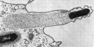
Rather than targeting metabolic enzymes, the current strategies being explored to combat dormant bacteria target either the membrane, or membrane bound proteins. Both of these approaches destabilise the bacterial membrane and help to break the cell apart and can act against processes such as energy synthesis which occur in both active and dormant cells.

a=targeting important metabolic proteins in the membrane. b=targeting the actual cell-membrane. Picture is copywrite me :p

In eukaryotic cells, such as the cells of plants and animals, the enzymes that create energy for the cell are kept safely hidden away in specialised intracellular compartments, such as mitochondria. As energy production requires an ion gradient across a membrane, these compartments all have sets of internal membranes. Bacteria however do not have this luxury, and instead have all their metabolic enzymes in the outer cell membrane, as this is the only membrane they have. Inhibitors of energy metabolism can therefore bind directly to target enzymes in the membrane involved in the production of energy. This can be highly effective against cells whose interior is hard to get into, such as Mycobacterium tuberculosis which lurks inside tuberculosis granulomas. Even in the absence of growth, cells still require a minimal energy input to survive, so blocking off these enzymes kills both dormant and active cells.

Drug developed to help combat TB by attacking cell membrane metabolic enzymes. This drug is currently in stage three clinical trials.

The membrane-targeting drugs act directly on the lipid bilayer that surrounds the bacterial cell, breaking it up and destroying the bacterial cellular integrity. Although human cells are also surrounded by lipid bilayers they have fewer negatively charged phosopholipids and also contain cholesterol (not present in bacterial membranes) allowing membrane-targeted drugs to be specific for human pathogens rather than killing surrounding human cells. The drugs that are used to attack the cell wall can vary hugely in size and structure but they all share one common property; they are highly lipophilic (i.e they are attracted to lipids). This allows them to interact with the cell membrane and break it apart.
Lipophilic drug capible of targeting bacterial cell membranes

There’s something about those molecular diagrams of drugs that I love. I think it’s my biochemical background. I’m never totally happy with a schematic until I can see how the chemicals are interacting on a molecular scale.

As well as being useful against dormant bacteria these new antimicrobials show promise as strategies for dealing with arising antibiotic resistance. Bacteria can evolve to cope with as many challenges as are thrown at them, but hopefully it should take them a little longer learn to survive entirely without a cell wall…

Although there are some that can do that already.

---
Hurdle JG, O'Neill AJ, Chopra I, & Lee RE (2011). Targeting bacterial membrane function: an underexploited mechanism for treating persistent infections. Nature reviews. Microbiology, 9 (1), 62-75 PMID: 21164535

---
Follow me on Twitter!

Bacterial comet tails

ResearchBlogging.orgI haven't worked very much with bacteria that infect humans. Most of my lab work has been done in the fields of either synthetic biology (which works with model organisms) or antibiotic production, which works on soil bacteria that produce the antibiotics. Human bacterial parasites therefore hold the fascination of the slightly exotic, not least because they sometimes do things like this:

Figure from"Molecular Biology of the Cell, Fourth Edition"by Alberts et al.

I've written before about some of the interesting features of intracellular bacteria, but this is possible one of the more exciting and fun things that they do. The picture above shows a eukaryote (human) cell outlined as a green oblong. Within the cell are lots of invasive bacteria (the red dots) some of which have a beautifully long green 'comet tail' flying out behind them.

That comet tail isn't just for show, it is vitally important for movement. The inside of a eukaryote cell is a fairly crowded and busy place, bacteria can't just swim around inside the cell like they would in the wild. Instead they have to rely on physical methods to push them through the cell and like invading virus's (which I wrote about here) they hijack machinery inside the cell to move them around.

Virus particles can latch onto the intracellular transportation machinery to hitch a free ride, but bacteria are too big for that. Instead, what most of them do is to produce proteins known as nucleation-promoting factors. These co-opt cellular proteins (the Arp2/3 complex for anyone with a background in actin polymerisation) which form branched actin fibres behind the bacterial cell, pushing it forward. The 'comet tail' pattern seem above, is seen by using a green stain for the structural actin protein, so you can see it forming long fibrous complexes behind the bacteria. These actin tails can move the bacteria wherever they want to go in the cell, and can also help with the invasion of neighbouring cells. This process is shown diagrammatically below:

Figure from"Molecular Biology of the Cell, Fourth Edition"by Alberts et al.

The actin system is a good one to use, as actin is ubiquitous inside all eukaryotic cells. In normal cell conditions is it used for structural purposes and is vital in cell division. Also important for the pathogenic bacteria (which after all does not want to kill its host straight away) is that none of these important cellular processes are compromised by the bacteria 'borrowing' some of the actin to move around with.

Another interesting point is that different intracellular bacteria often produce different types of actin tails. L. monocytogenes and S. flexneri have short, highly crosslinked filaments producing short stubby little tails, whereas Rickettsia species have actin tails that are composed of distinctly longer bundles of unbranched actin filaments. Part of the reason for this is that different bacteria will produce different nucleation-promoting factors and some of the more lazy ones (i.e S. flexneri) don't even bother to do that and just use the host nucleation-promoting factors within the invaded cell! Recent work has shown that Rickettsia on the other hand, doesn't even rely on the host Arp2/3 complex to polymerase the actin and instead relies almost entirely on their own, bacterial, proteins.

They truly are beautiful to look at though. Even without all the fancy colour staining:


Listeria monocytogenes pushing right at the cell membrane, with actin tail behind. Electron micrograph picture taken from the se reference below

---
Ray K, Marteyn B, Sansonetti PJ, & Tang CM (2009). Life on the inside: the intracellular lifestyle of cytosolic bacteria. Nature reviews. Microbiology, 7 (5), 333-40 PMID: 19369949

Kuo SC, & McGrath JL (2000). Steps and fluctuations of Listeria monocytogenes during actin-based motility. Nature, 407 (6807), 1026-9 PMID: 11069185
---
Follow me on Twitter!

Hitchhiking through the nervous system

ResearchBlogging.orgI while ago I wrote a post about how virus's get from the outside of the cell to the interior of the nucleus and found that virus particles are able to hitchhike on the cells internal transport systems. I was quite interested therefore to find a paper in Nature Reviews (reference below) that revealed that not only do virus's latch on to host proteins to travel around inside the cell, they also use host extracellular processes for travelling around the body. And outside the cell it's not just virus's either, bacterial toxins need transport systems too, unlike whole bacteria they can't move around under their own power.

One place that the body wants to protect particularly well against infection is the central nervous system. It provides this protection by surrounding it with a wall of tightly sealed endothelial cells known as the blood-brain barrier. However despite this the body itself still need to get some things into the CNS; small molecules such as glucose and oxygen as well as larger cells of the immune system. These immune system cells provide the first sneaky point of entry; virus's such as HIV can hitch a ride inside these cells and get into the central nervous system that way. This is the equivalent of hiding in a truck to avoid border patrols.

However some virus's and toxins use an even more sneaky method, dressing up as a border-patrol guard and simply walking in. Throughout the blood brain barrier there are long neuronal projections that connect the central nervous system to peripheral organs. A picture of one of these cells is shown below:

Like all cells, this contains the transport molecules Kinesin and Dynein, which virus's can latch onto in order to transport themselves through the cell (see earlier post here). Once they get inside the cell, the cell's own proteins will carry the virus particles all the way through it, and into the central nervous system. However first it has to get inside the cell, through the little blue blob at the bottom (in the diagram above it's highlighted with a little dotted square).

As well as receiving chemical signals for electrical impulses (that make the neuron function as a nerve) the blue blob also contains various different receptors capable of engulfing and uptaking small molecules, including those used to signal some neural impulses. This means that there are a range of chemical receptors on that blue blob which allow the uptake of molecules, and you can probably tell where this is headed...
The diagram above is the intramolecular equivalent of Han Solo dressed as a Stormtrooper wandering into the Death Star. By changing its outer coat enough to mimic the proteins that are usually taken up by the cell the Herpesvirus can attach to the outer membrane and then be absorbed into the cell. Once inside, it can latch onto the dynein and get a free pass all the way into the nucleus (and neurons are pretty long so it is a bit of a journey). Poliovirus and rabies can also carry out this trick (at the neuromuscular junction for anyone interested) along with the bacterial botulinum toxin, which gets taken up by synaptic vesicles and essentially kills the end of the nerve, which can either lead to instant death or a scarily smooth robot-plastic forehead, depending what context you take it.

I always find it quite spooky to think of my body in that way, as a huge maze of intracellular processes, being negotiated, infected and protected by tiny substances outside of my conscious control. I think that's another reason I find cellular biology so fascinating, by studying it we gain control (or if not control at least an understanding) of these detailed processes that we would not normally be able to influence.

---

Salinas S, Schiavo G, & Kremer EJ (2010). A hitchhiker's guide to the nervous system: the complex journey of viruses and toxins. Nature reviews. Microbiology, 8 (9), 645-55 PMID: 20706281

Bacterial needles and their role in infection

This post was chosen as an Editor's Selection for ResearchBlogging.orgI spent ages over the title of this post. The original paper "Injecting for infection" was my favorite but it isn't wonderfully clear. It sounds like something about dirty needles; bacterial colonies over the surface of injections. In reality it's about something far more amazing, the little needles that bacteria make themselves, in order to inject toxins into the cells that they destroy.

Officially these are called Type III secretion systems, as they allow the secretion of toxins (and other things for that matter) from the cell. They occasionally fall off the bacteria allowing very detailed electron microscope pictures to be taken, showing that, whatever their official name, they do look a lot like little needles.

Image A shows the imprint of the needle on the bacterial cell surface. Image B shows the isolated needles, showing their structures (which are wonderfully detailed and quite beautiful). Image C shows a drawing of the proteins involved in the structure within the cell membrane. Scale bar is 100nm.

In infectious organisms this secretion system is vital for survival, but it's interesting to see how it's used in organisms that are only opportunistically infective, such as Pseudomonas aeruginosa. P aeruginosa is an opportunistic pathogen, it can survive fine outside the human body, but in cases where it gets a chance to invade (particularly in the lungs of people with cystic fibrosis) it will go for it. Where it inherited the needle complex from is not clear, although it is thought to have distantly evolved from flagella and been passed to the pseudomonas by horizontal gene transfer from another bacteria.

Removal of the needle complex does not prevent P aeruginosa from invading and infecting an organism, but it does make the infection slightly less virulent. Work on acute pneumonia has started to build up a model of how the needle works, and what role it plays in infection. The bacterial cells invade the epithelial tissue in the human host at points where it is damaged (i.e by cystic fibrosis). As non-damaged epithelial cells are usually quite resistant to the bacterial colonization, it is only when the lung tissue is already injured that the Pseudomonas can take hold.

Once P. aeruginosa has colonized the damaged tissue surrounding macrophages and neutrophils will gather at the site of infection. These merely further damage the surrounding tissue, without harming the bacteria allowing it to settle and grow. Only then does the needle start pumping out damaging toxins, which lead to the symptoms of pneumonia. In a severe infection this can lead to a breach of the tissue barrier between the lungs and the blood stream, which goes on to cause systemic bacteria infection and rapid septic shock. The removal of the needle complex can therefore stop some of these more extreme reactions, but does not prevent the infection starting in the first place.

---
Hauser, A. (2009). The type III secretion system of Pseudomonas aeruginosa: infection by injection Nature Reviews Microbiology, 7 (9), 654-665 DOI: 10.1038/nrmicro2199