Field of Science

Models for the evolution of bacterial resistance

This post was chosen as an Editor's Selection for ResearchBlogging.orgI wrote about Quorum Sensing recently, the ability of bacteria to communicate with each other via small molecules which they can both excrete and sense. A lot of the research done on quorum sensing aims to find ways to block the system, as it is one of the main communication methods used to switch on virulence genes, and other genes which make the bacteria more infectious, and more likely to cause harm.

Blocking quorum sensing would not in theory kill the bacteria, but it would give more time for the bodies immune system to recognize and destroy the bacteria. This is starting to be a more common approach in the design of anti-microbials, and in virus vaccines. A slower and less virulent form of both virus's and bacteria can sometimes be dealt with easily enough by the body's own immune system as it has more time to respond to the invading challenge.

There is also an evolutionary reason for the effectiveness of quorum sensing disruption over conventional antibiotics. Antibiotics kill off any bacteria that aren't resistant, creating a highly competitive growth environment with a large selection bias against non-resistant bacteria. Disrupting the quorum sensing system on the other hand should mean that the bacteria are still growing in a non-selecting environment, right up to the point where the body's defense system kills them off. There is less evolutionary pressure on them to develop resistance.

A paper in PLoS (reference below) looked at developing a method for how (and whether) resistance towards quorum sensing disruption might occur. They chose to base their model on the effects of altering the genes involved in quorum sensing, rather than other methods for quorum sensing disruption, such as blocking or destroying the little molecules used for signalling and one of the first things they found was that there are a large number of variations in these genes, despite the fact that many of the actual signalling molecules are fairly similar.

Lots of gene variation means bad news for gene-based disruption as there is potential for the bacteria to share these genes among themselves, and thus potentially end up with a combination that renders the gene-blocking anti-bacterial invalid. And swopping genes is one of the things bacteria do best.
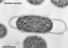
Figure above shows two bacteria connecting together via a long thread-like molecule called a 'pilus'. The genes do not actually travel through the pilus, but the pilus pulls both bacteria close together allowing the gene (in the form of a plasmid) to be passed between them.

Another thing that was examined was the effect of quorum sensing disruption on fitness. The idea that resistance to quorum sensing (which will henceforth be called QS as I can't be bothered to keep typing it out) will take more time to develop is based heavily on the idea that tampering with the QS does not affect the general health of the bacteria. Sure it prevents bacteria from communicating, but each individual bacteria is no less likely to thrive, and therefore there is less selection pressure.

However almost all of these experiments (certainly those in academic labs) have been done in conditions of nice safe uber-rich bacterial growth media. Even slightly shaken bacteria may thrive fine on nutrient rich agar. Inside an actual living organism though (the normal living place of pathogenic bacteria) disrupting the QS system has major implications. Not just in the fact that non QS-able organisms are often outcompeted by other bacteria, but also because some QS-controlled proteins seem (under certain conditions) to be vital for bacterial growth. If disruption of QS is leading to bacterial death then this puts a massive evolutionary pressure on bacteria to evolve resistance, similar to the pressure to evolve antibiotic resistance.

Interestingly (if slightly off topic) it is vaguely suggested that the necessity of QS for growth might help to prevent 'cheaters' within a bacterial population; bacteria that take up all the benefits of it's QS-ing neighbors (i.e biofilm formation, increased virulence etc) without having to expend energy into the QS system.

In view of this data it looks as if, despite it's undoubtable advantages as an antimicrobial, QS-disrupting systems will inevitably succumb to bacterial resistance. In the presence of a selective pressure favoring it resistance will always develop. There is no golden bullet for antibacterial strategies, just a lot of little silver bullets that eventually loose their power.

So as not to end this on a depressing note, it is worth mentioning that the fact that there is a lot of research going into alternative (i.e not antibiotics) antimicrobials is encouraging. Phage therapy and QS disruption will probably never replace antibiotic therapy, but it is worth having as many different strategies as possible to deal with the threat of bacterial infection.

---
Defoirdt T, Boon N, & Bossier P (2010). Can bacteria evolve resistance to quorum sensing disruption? PLoS pathogens, 6 (7) PMID: 20628566

---
Follow me on Twitter!

Cherry Beer and Carnivals

I've just come back from a quick weekend away in Brussels which was really amazing. I would definitely recommend it as a holiday destination, it's easy enough to get to by Eurostar and there's plenty to do there. We went to the Instrumental Museum and the Cinema Museum (with clips from the first ever films) ate lots of mussels and interesting seafood and I discovered Kriek:


I'm not really a beer drinker, but Kriek is a form of cherry beer, which is really pleasant. Sitting in the sun outside a cafe, with Kriek and mussels talking with my fiancé about ancient cinema clips (including some really early Disney stuff) was just amazing :) It was a great holiday, and seemed far longer than just three days sneaky break.

I was going to try and write a science-post but I've been working double-speed today to make up for taking the day off so unfortunately new bacteria knowledge won't be materialising until later in the week. I would encourage anyone who has any interesting molecular biology posts to submit them to the Molecular Biology carnival though, not long until the first every issue and it would be great to make it a nice full one! Submit here, or by using the widget on the sidebar.

Quorum Sensing and Biofilms

This post was chosen as an Editor's Selection for ResearchBlogging.orgAlthough bacteria live as isolated cells, they are constantly communicating with surrounding bacteria, particularly those of the same species, which can often band together to form large groups of bacteria surrounded by a sticky mesh. These are known as biofilms (which I cover in more detail here). One of the main ways that bacteria communicate with each other in order to organise structures like this is by quorum sensing.

Quorum sensing uses small molecules that bacteria can both excrete and sense. When enough bacteria are in one place then the surrounding concentration of these small molecules reaches a critical level and can activate the genes for a variety of different responses including luminescence, virulence (in pathogenic or disease-causing bacteria) and the formation of biofilms:
A recent paper (reference below) looked at a range of different types of biofilm and quorum sensing interactions, in order to explore the different environmental pressures that shaped the differences in these systems. They found that although many species formed biofilms when bany cells joined together some species stopped forming biofilms when they reached a certain cell density. Biofilms are carefully controlled by bacteria, they do not just start growing when a certain number of bacterial cells gather together and then never stop.

Using models of mostly infectious biofilm-forming bacteria (such as Vibrio cholerae which causes cholera) they found that as well as helping to bind the cells together and resist man-made antibiotics (which cannot penetrate the biofilm) the biofilm was also a defense against competing bacteria (and may have helped to out-compete them by covering all available living surfaces with slime). The ability to produce biofilms not only helps the V. cholerae against other invading bacteria, it also helps it gain a hold against the body's own internal bacterial defenses that line the internal gut.

However once the levels of V. cholerae became too high the bacteria often stopped generating the biofilms. This could be for two reasons, firstly the biofilm takes up valuable resources that could be used in growth and division and secondly it prevents the bacteria within it from travelling very far. V. cholerae infect the body by having periods of growth followed by periods of mad colonisation, which works best if the biofilm actually disperses at high cellular density to allow the cells to spread.

This can be contrasted with more sedentary bacteria like P. aeruginosa which likes to settle down once it finds a place to live and occasionally disperse colonies into the body. Rather than loosing its biofilm this bacterial species retains it even at high cell densities. This allows it to out-compete any other bacteria that may be at the site of infection, and hold off both the body's natural defenses and any chemical antibiotic drugs meant to kill it.

Comparisons of different V. cholerae strains revealed a wide range of different biofilm formation patterns between strains, all linked to Quorum sensing signalling. This is likely to depend on the internal environment that specific strains occupy, the amount of competition they face and the necessity for quick and frequent bouts of dispersal.

As biofilms are traditionally studied in P. aeruginosa I found it fascinating to hear about how other bacteria use them in order to colonize their surroundings. In aeruginosa biofilms are a mark of stability, the bacteria have found a place to stay and invest time and energy in making it as safe and indestructible as possible. In cholerae however, the biofilm is just protection for the growing bacteria, until their numbers get high enough to allow them to break out and invade the body.

---
Nadell CD, Xavier JB, Levin SA, & Foster KR (2008). The evolution of quorum sensing in bacterial biofilms. PLoS biology, 6 (1) PMID: 18232735

How viruses hijack cellular transport systems

ResearchBlogging.orgEven in the world of the very small, there are significant differences in size. A eukaryote cell (i.e a human cell) for example is relatively big, in microscopic terms. Most other things that interact with the cell at the microscopic level, are far smaller than it, such as bateria, viruses and signalling molecules.

A virus isn't much more than a small capsule of proteins with a little bit of DNA inside. Once it gets inside a eukaryote cell, it's very much in the position of a small child wandering into a big city. In front of it lies the vast interior of the cell, full of reactions, enzymes, proteins scurrying too and fro, mRNA being translated, proteins being folded and other busy bustling cellular processes. Surrounding it are large organelles (larger than the virus particle!) with strange and mysterious procedures going on inside them...

That sort of view, stretching across to both horizons

From here, the virus has to make its way to the nucleus, pushing its way through the crowded and complex cellular interior without being spotted as an intruder. Fortunately it has some help here, because it's facing the same problem faced by every molecule and organelle already in the cell. Transport mechanisms are already in place so that things can move around the large intracellular space with relative ease. The viruses simply hijack these transport systems and get a free ride all the way too the nucleus.

Work on the herpes simplex virus helped to produce a model of how the viral particles move around the cell. After entering the cell through the cell surface membrane, the virus is picked up by dynein which carries it along microtubes towards the nucleus. The microtubules form a network within the cell (like train rails) which the dynein motors along (using ATP energy). This is shown pictorally below:

Dynein moves in one direction along the microtubule while kinesin moves in the other direction. Together they move molecules all around the cell.

Once at the nucleus, the viral DNA enters through the nuclear membrane and is replicated inside the nucleus (entering the nucleus is a critical step for DNA-viruses; for those viruses that contain RNA this step is not so vital). The replicated DNA then comes back out of the nucleus and is transcribed into protein in the cytoplasm, which leads to the formation of new viral particles. These new viruses then have to travel back down the microtubule (carried by kinesin) to the outer membrane of the cell where they can be released into the surrounding environment and go on to infect more cells.

One interesting question is what exactly the dynein (and kenesin) bind to on the virus cell surface. As well as being an interesting point, answering this comes with the usual funding bait that if you find how viruses move inside the cell you may be able to find ways of stopping them from moving which would leave them at a severe disadvantage. To examine this the virus was isolated and the parts of the surrounding protein coat that bound to cellular factors further separated. These separated capsid proteins were then tested for their ability to bind to mammalian intracellular proteins. They found that several of the capsid proteins could bind to important transporter molecules, and furthermore that several different transporter molecules could sometimes bind to the same capsid protein.

Drawing showing the site of attachment of the motor transport proteins to the (green) virus capsule. The other end of the motor proteins is used to move along the microtubule.

As I'm in a fairly syntheticly-biological mood, I couldn't help but notice the mention at the end of the paper that this could have implications beyond virus treatment or vaccinations. The ability to create a little molecule that the cell can carry to the nucleus could have implications for both future genetic treatments and nanotechnology. The ability to get a little capsule of treatment right to the nucleus of cells could even have the potential for treating cancer cells, as it utilizes the cells own transport mechanisms to deliver treatment to the intracellular place it is needed.

---

Kerstin Radtke, Daniela Kieneke, André Wolfstein, Kathrin Michael, Walter Steffen, Tim Scholz, Axel Karger, Beate Sodeik (2010). Plus- and Minus-End Directed Microtubule Motors Bind Simultaneously to Herpes Simplex Virus Capsids Using Different Inner Tegument Structures PLoS Patholgens, 6 (7) : e1000991

---

Follow me on Twitter!

Bacterial needles and their role in infection

This post was chosen as an Editor's Selection for ResearchBlogging.orgI spent ages over the title of this post. The original paper "Injecting for infection" was my favorite but it isn't wonderfully clear. It sounds like something about dirty needles; bacterial colonies over the surface of injections. In reality it's about something far more amazing, the little needles that bacteria make themselves, in order to inject toxins into the cells that they destroy.

Officially these are called Type III secretion systems, as they allow the secretion of toxins (and other things for that matter) from the cell. They occasionally fall off the bacteria allowing very detailed electron microscope pictures to be taken, showing that, whatever their official name, they do look a lot like little needles.

Image A shows the imprint of the needle on the bacterial cell surface. Image B shows the isolated needles, showing their structures (which are wonderfully detailed and quite beautiful). Image C shows a drawing of the proteins involved in the structure within the cell membrane. Scale bar is 100nm.

In infectious organisms this secretion system is vital for survival, but it's interesting to see how it's used in organisms that are only opportunistically infective, such as Pseudomonas aeruginosa. P aeruginosa is an opportunistic pathogen, it can survive fine outside the human body, but in cases where it gets a chance to invade (particularly in the lungs of people with cystic fibrosis) it will go for it. Where it inherited the needle complex from is not clear, although it is thought to have distantly evolved from flagella and been passed to the pseudomonas by horizontal gene transfer from another bacteria.

Removal of the needle complex does not prevent P aeruginosa from invading and infecting an organism, but it does make the infection slightly less virulent. Work on acute pneumonia has started to build up a model of how the needle works, and what role it plays in infection. The bacterial cells invade the epithelial tissue in the human host at points where it is damaged (i.e by cystic fibrosis). As non-damaged epithelial cells are usually quite resistant to the bacterial colonization, it is only when the lung tissue is already injured that the Pseudomonas can take hold.

Once P. aeruginosa has colonized the damaged tissue surrounding macrophages and neutrophils will gather at the site of infection. These merely further damage the surrounding tissue, without harming the bacteria allowing it to settle and grow. Only then does the needle start pumping out damaging toxins, which lead to the symptoms of pneumonia. In a severe infection this can lead to a breach of the tissue barrier between the lungs and the blood stream, which goes on to cause systemic bacteria infection and rapid septic shock. The removal of the needle complex can therefore stop some of these more extreme reactions, but does not prevent the infection starting in the first place.

---
Hauser, A. (2009). The type III secretion system of Pseudomonas aeruginosa: infection by injection Nature Reviews Microbiology, 7 (9), 654-665 DOI: 10.1038/nrmicro2199

Programming bacteria for search and destroy

ResearchBlogging.orgAs iGEM season is now properly underway, I thought I'd have a look at a synthetic biology paper and found this fairly awesome one about programming bacteria to hunt out and destroy atrazine, a chemical herbicide pollutant. One of the most exciting things about this work was that it didn't just involve bacteria with the ability to remove atrazine from the environment but to actively migrate towards the chemical and then destroy it.
The chemical structure of atrazine

The bacteria are controlled using riboswitches - little RNA pieces that can bind directly to a ligand (or signal molecule) and cause a change in gene expression, changes which can include switching on or off the genes involved in cell movement. Atrazine is a good molecule to start with because as well as being a relevant pollutant it also contains plenty of N-H bonds which are good for forming hydrogen-bond interactions with RNA. As well as that it has a well-characterized breakdown pathway, all components of which have been expressed successfully in E. coli.

The first stage in creating these seek and destroy bacteria was finding RNA sequences that would bind to atrazine. This was done by attaching the atrazine to a solid support and running bits of RNA past it, to see which ones would bind. They then took these successful binders and tested for riboswitch activity, i.e whether the binding to atrazine caused a conformational change in the RNA that lead to the turning on of a gene. They did this by putting a sequence complimentary to the isolated RNA upstream of the DNA for the CheZ gene, which controls motility in E. coli and then carrying out the selection process shown below:

In the absence of atrazine the CheZ is not synthesized and the bacteria stay where they are. When atrazine is added to the plate, the bacteria start to move...

Dose-dependent assays were then done on the successful RNA sequences, to characterise the reaction and check that it actually was the atrazine levels that lead to movement rather than some other confounding factor. Sequencing and examination of the binding site also helped to characterize the riboswitch and determine how it was working. The genes for atrazine-consuming ability were then added to the bacteria that moved towards the atrazine, leading to a little search-and destroy module capable of seeking out a dangerous pollutant and removing it from the environment.

---
Sinha J, Reyes SJ, & Gallivan JP (2010). Reprogramming bacteria to seek and destroy an herbicide. Nature chemical biology, 6 (6), 464-70 PMID: 20453864

---
Follow me on Twitter!

New Carnival!

This is a little late as I've been away, but better late than never! A new carnival has arrived thanks mostly to the efforts of Alejandro Montenegro aided by myself, Lucas Brouwers, Psi Wavefunction and Alexander Knoll.

This carnival will collect articles focussing on all processes that go on inside cells. Every month (starting August the second) a collection of these articles will be gathered and posted on a suitable host blog. Posts from anyone are welcomed, as long as they feature internal cellular happenings.

I really love blog carnivals, as they give an interesting selection of work to browse through and, from a more selfish perspective, they do help to increase readership to your blog. Submissions will be accepted up too the first of August so if you have written, or are planning to write, anything about the Private Life of Cells then go submit your post here.

Also, for those that missed it the latest Carnival of Evolution is up at Culturing Science. It's an absolutely amazing issue, with loads of great articles and little hand drawn illustrations for each section heading.

Life at 90°C

ResearchBlogging.org
Prokaryotes are by far the most successful superkingdom in terms of types of both biochemical diversity and the variety of environments conquered. Bacteria can be found living in all kinds of adverse conditions; from high alkaline lakes, to below freezing temperature, to hot volcanic vents which in some cases can reach temperatures close to the boiling point of water.

Thermotoga is a small genus of bacteria that contains some of the most hyperthermophilic species known, some able to survive at 90°C although most prefer the cooler temperatures of 70-80°C. It’s called “thermotoga” because it lives at high temperatures (thermo) and contains a characteristic outer cell membrane known as the ‘toga’.

This was basically my mental image when I read that

One of the most interesting things about thermophilic bacteria (i.e bacteria that like living at very high temperatures) is their enzymes. Most normal enzymes, (most normal proteins in fact!) will break down and denature at very high temperatures, so bacteria like thermotoga will usually have their own set of enzymes. These enzymes are usually of great interest to people carrying out industrial processes, which all take place at higher temperatures. The most important enzyme in the PCR protocol (Taq polymerase, which synthesises DNA at temperatures of up to 80°C) was isolated from a marine thermophilic archaea.

Another exciting thing about Thermotoga is it has the ability to produce hydrogen. In the lab, it uses carbohydrates from yeast extract or peptone to form sugars, which are oxidised to carbon dioxide (or acetic acid) using either sulphur or protons as the electron acceptor. Where protons are used, they get reduced to hydrogen as the carbon dioxide is formed. The yield of hydrogen from glucose is very high, and in many cases can approach the theoretical maximum yield of for 4 mole hydrogen from one mole off glucose. This represents almost twice the amount that can be obtained from other bacterial hydrogen producers. Because this process is taking place at high temperatures, the enzymes involved in the process could potentially be used inside high temperature reactors.

Thermotoga neopolitana has the potential to produce one of the highest hydrogen yields of all as it is able to respire microaerobically. This means that it would be theoretically possible for the cells to aerobically oxidise a small amount of the glucose, generating enough energy to ensure that all the remaining glucose is fully oxidised by hydrogen-generating pathways. As yet, the metabolic pathways for oxygen use within these organisms have not been identified, but it does look as if hydrogen generation depends almost exclusively on anaerobic processes.

Electron micrograph of Thermotoga, showing the large outer cell wall covering (the toga!).

As the majority of the research into hydrogen production of Thermotoga has focussed on yield, issues of productivity, stability and required substrates will all need to be address in order for this process to be fully understood and possibly implemented in an industrial setting. There are also issues with biomass concentration – which is restricted, possibly by free-flowing biofilms (and possibly due to the toga-like nature of the surrounding cell-wall capsules. However even if large scale hydrogen production proves to be unfeasible with these microorganisms, studies of the enzymatic processes used to produce hydrogen at high temperatures may have applications above and beyond using the entire bacteria for hydrogen production.

---

Van Ooteghem, S., Beer, S., & Yue, P. (2002). Hydrogen Production by the Thermophilic Bacterium Thermotoga neapolitana Applied Biochemistry and Biotechnology, 98-100 (1-9), 177-190 DOI: 10.1385/ABAB:98-100:1-9:177