Field of Science

Getting OMPs to the membrane - SGM series

ResearchBlogging.org This is the first post of my SGM conference series: I'm going to try and write about seven topics from the Society for General Microbiology September conference over the course of two weeks. The first topic I'm looking at is Protein Folding and Misfolding which consisted of thirteen presentations covering various aspects of protein folding in bacteria, fungi and yeast. As a quick background: when proteins are synthesized they are constructed as long chains of amino-acids which then need to fold up into the correct shape.

This may not sound terribly interesting at first, but it proves problematic for proteins that are in awkward places, for example in the outer membrane of Gram negative bacteria. These outer membrane proteins (OMPs) not only have to fold up correctly inside the membrane but they have to actually get to the outer membrane In Gram negative bacteria, this means first getting through the inner membrane, then across the peptidoglycan layer between the membranes, and finally half way through the outer membrane in order to coil up correctly inside it.

Gram negative cell membrane

The type of proteins found inside the outer membrane are usually B-barrel proteins, so called because they contain lots of protein folds known as a B-sheets, which can wrap up to form a channel shape as shown in the example on the right. Each blue arrow is a single B-sheet and these fold specifically to form pore-like structures which in the case of porins make a little hole through the membrane.

Transport of B-barrel OMPs accross the inner membrane is achieved by synthesising them with a signal sequence attached to one end. This signal sequence is recognised by proteins on the inner membrane and ATP energy is used to pump the proteins accross the inner membrane and into the periplasm (the space between the two membranes). Once in the periplasm they bind to little chaperone proteins which carry them safely to the complex responsible for folding them correctly into the outer membrane, the rather awesomely named BAM complex.As an aside the chaperones do have to get the OMPs there fairly promptly as there are proteases that float around in the periplasmic space and degrade any proteins that don't get incorporated into the outer membrane quick enough.

One of the key proteins in the BAM complex is BamA as knocking it out results in a lot of unfolded OMPs in the periplasm (and probably a field day for the proteases). BamA consists of two major components, a B-barrel domain which anchors it into the outer membrane, and five "polypeptide transport-associated" domains, shortened to POTRA by someone who didn't like three-lettered acronyms. The POTRA domains do what they say, they are associated with the transport of proteins (polypeptides).

It's still a little uncertain quite how the BAM complex works but a couple of the presentations on the topic were convering it, including work done on changing the genes between different bacterial species. All Gram-negative bacteria have a BamA gene, however taking the BamA gene from one bacteria and putting it into another does not end happily unless it's done between two very close species. Closer research with chimeric proteins (i.e proteins that are half from one bacteria and half from another) shows that this only applies to the POTRA domains. The anchoring B-barrel can be switched between several different species, but the POTRA domain is very species specific.

Another interesting thing to address is how BamA gets itself into the outer membrane. One of the periplasmic chaperone proteins, Skp, is thought to be involved in this process, and it was found that when the outer membrane was negatively charged Skp is involved in inserting BamA into the membrane, whereas when the negative charge is removed Skp inhibits BamA folding and insertion. Negative charge is caused by an increase in the phosphatidylglycerol content in the membrane. I found that idea quite exciting as it implies that the bacteria can control where they want the BAM complex to go. The idea of membranes forming "lipid rafts" with certain components that organise where proteins are held is not a new one, and the BAM complex forming in specific places in order to create the correct outer membrane protein concentration is one that appeals to me.

They may just be single little cells with no true nucleus, but they are capible of a lot of control over their intracellular processes!

---
Knowles TJ, Scott-Tucker A, Overduin M, & Henderson IR (2009). Membrane protein architects: the role of the BAM complex in outer membrane protein assembly. Nature reviews. Microbiology, 7 (3), 206-14 PMID: 19182809

Johnson, A., & Jensen, R. (2004). Barreling through the membrane Nature Structural & Molecular Biology, 11 (2), 113-114 DOI: 10.1038/nsmb0204-113
---

Follow me on Twitter!

Unfortunate change of plans

So...anyone following me on Twitter probably saw me getting all excited when I signed up to go to the Society of General Microbiology conference a couple of months ago. The conference is next week, but unfortunately I won't be at it, which is a pity because I was all set to blog about it and everything.

There are two main reasons that I can't make it:

1-Time. I'm just finishing off my summer project and starting to need/get exciting results. Also I'm sorting out my wedding, trying to get a PhD organised, and heading off to another more work-related conference later this month. Taking four days off would be slightly overindulgent at this point.

2-Money. I do not have any. As an undergraduate SGM member I can't get a travel grant from them (graduates only) and as I've graduated the university isn't about to give me any money. So I have to pay to transport myself up there, and for somewhere to sleep for four nights. Train fares are expensive nowadays and even the cheapest B-and-B I had planned came to £180. Quite frankly if I had £180 I'd use it to clear my overdraft.

HOWEVER - all is not lost. As I registered for going to the conference (for free - the perks of being an undergraduate member) they sent me a PDF of the abstracts for each session. So, over the next two weeks I'm going to have a rather sad and nerdy little single-person conference of my own on my blog. I'm going to try and get a post up every other day (so seven posts over two weeks) covering one topic at the conference with each post.

I've got a choice of seventeen, but here are the topics I'll probably aim to cover as they're the ones I most wanted to see:

Metals and Microbes
Streptococci
Acid Stress
Microbial Death
New Insights into Secondary Metabolism
Extremophiles
Protein Folding and Misfolding
(and if I have time, Microbial Models of Human Diseases)

Before anyone starts feeling too sorry for me about missing this I should point out that a) I have another conference I'm going to this month b) that conference is fully funded and c) that conference is in Italy...

And there will be many more conferences in my life, I'm sure!

Trojan horse predators

This post was chosen as an Editor's Selection for ResearchBlogging.org A while ago, Angry by Choice wrote a post about a fungi that kills its nematode prey by making little lasso ropes to catch the worm in. At the time, I thought there must be some exciting way that bacteria could cause wormy destruction, but it wasn't I read a paper from Lucas (reference below) that I actually found one.

It's not as visually exciting as the little fungi nooses, but it's just as chemically exciting. As bacteria are not capable of forming phyiscal structures to capture a worm, they make chemical ones, specifically volatile chemicals which diffuse easily and can be sensed by the worm.

In layman terms they smell good. They smell like food.

This is officially the best MS Paint picture I've ever done

Nematodes don't have a huge number of well programmed behaviours in their little nematode brains, but "move towards food" aka "positive olfactory chemotaxis" is one of the most robust and common behaviors. And the bacteria take full advantage of this. They secrete chemicals that are based on modified quorum sensing molecules (usually used for bacterial communication), which cause the worms to not only arrive where the bacteria are waiting, but also to happily gobble them up, after all they do smell like food.

Once eaten, the bacteria end up in the worms digestive tract, where they start to secrete enzymes - two digestive proteases called Bace16 and Bae16. Although previous work had assumed that these two proteases worked on the outside of the worms cuticle the paper used flourescent labeling studies to show that both Bace16 and Bae16 had their effects once inside the worm. Bace16 (labelled in red) and Bae16 (labelled in green) were both injected into an unsuspecting worm, which was then visualised every hour:

Images taken after 2hs, 5hrs, 8hrs and 24hrs - from the reference.

It's a little hard to see in the small picture above, but it is clear that the worm is getting ill, breaking apart, and finally just decomposing due to the action of the two proteases. The bacteria is not using the proteases to break into the worm, but to break out of it, digesting the worm in the process. This was further proved by making bacterial strains with the genes for Bace16 and Bae16 knocked out. Infection with the knockout strains led to far less virulent bacteria, and worms that survived for far longer.

It's an interesting new type of predation - a kind of Trojan Horse predator. Rather than chasing its prey, or directly infecting it, the bacteria gets itself eaten and then destroys the worm from the inside out. The paper suggests that as well as being interesting, this knowledge could help lead to more efficient biocontrol strategies for the elimination of nematode worms, now we know the active series of events involved in nematode predation by bacteria.

---
Niu Q, Huang X, Zhang L, Xu J, Yang D, Wei K, Niu X, An Z, Bennett JW, Zou C, Yang J, & Zhang KQ (2010). A Trojan horse mechanism of bacterial pathogenesis against nematodes. Proceedings of the National Academy of Sciences of the United States of America PMID: 20733068
---
Follow me on Twitter!

Nanofibre paint that kills MRSA

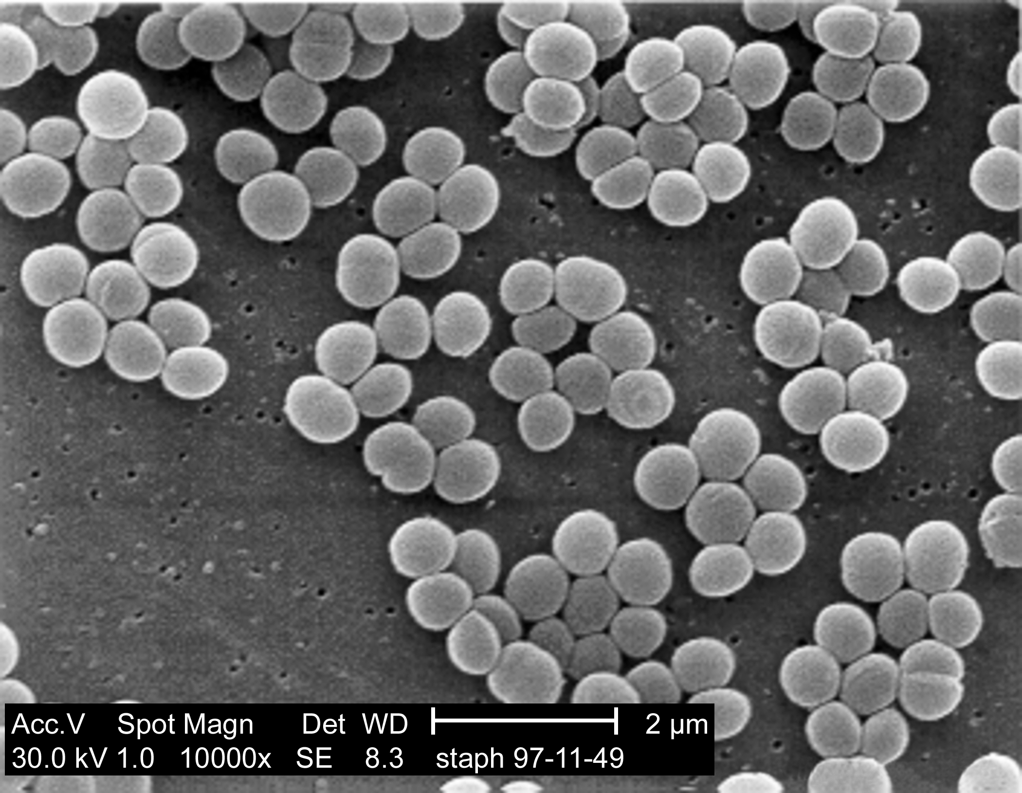
This post was chosen as an Editor's Selection for ResearchBlogging.orgMRSA, the antibiotic resistant form of Staphylococcus aureus is a major problem in hospitals. The antibiotic resistance makes it hard to erradicate, not just from patients, but in the surounding environment, on surfaces, on medical equipment, on the walls of the hospital. In order to minimise the numbers of dangerous bacteria found in hospital surroundings, quite a lot of research has gone into creating antibacterial coverings or coatings that would reduce the number of bacteria p. Currently however, many of these coating substaces work by either using biocides (such as silver) or releasing antibiotics and antimicrobials, which doesn't work on bacteria that have gained resistance.


Scanning electron microscope picture of clusters of Staph aureus

However medical scientists aren't the only ones trying to kill bacteria, virus's known as bacteriophages are also interested in breaking open bacterial cells and they do it using a cocktail of different enzymes to break open the cell wall. Many of these enzymes feature a two-domain structure with bacteria-specific cell wall targeting and catalytic domains. The enzymes Lst was found to be particularly good at breaking apart Staph aureus cell walls with one end of the enzyme (C terminus) recognising and binding to the bacterial cell wall while the other end (the N terminus) breaks the protein bridges between the sugar componants of the peptidoglycan layer, which is a major componant of the cell wall.

Schematic of the cell wall

Work from the Rensselaer’s Center for Biotechnology has been looking at incorporating these enzymes into nanofibres to create stable bactericidal paint films. The molecular-level curvature of carbon nanotubes stabilizes a wide range of enzymes and the lab was able to successfully create Lst-containing nanocomposite films which achieved >99.9% killing of MRSA upon contact within 2 h. They also explored incorporating these into a latex paint, which retained the bactericidal properties of the nanofibres. This paint could theoretically be spread over hospital surfaces to reduce the numbers of Staph aureus within the hospital environment. Incorporating this enzyme into the nanofibres (rather than directly mixing with the latex) gives added stability and helps the enzyme stay within the coating for longer. Films which were stored dry at room temperature showed >99% bactericidal activity against S. aureus after 30 days.

It remains to be seen how effective this technique will be outside of a laboratory setting but at the moment it looks like a highly promising step to help reduce the incidence of a dangerous pathogen. The speed and likelihood of resistance also remains to be seen, but it's heartening that bacteriophages have been using these enzymes against the bacteria for far longer than we've been using antibiotics. This is unlikely to be any kind of magical anti-MRSA cure, but it could certainly be very useful in helping to reduce the incidence of the disease.

---
Pangule RC, Brooks SJ, Dinu CZ, Bale SS, Salmon SL, Zhu G, Metzger DW, Kane RS, & Dordick JS (2010). Antistaphylococcal nanocomposite films based on enzyme-nanotube conjugates. ACS nano, 4 (7), 3993-4000 PMID: 20604574
---

Follow me on Twitter!

Art from Turing Patterns

Turing patterns are the more common name for "reaction-diffusion patterns" which are found in abundance throughout the natural world. They are formed by a simple system of cell-cell communication; cells secrete signals that mean nearby cells will become the same as them, whereas far away cells will differentiate. In terms of colour this leads to dots and stripes patterns, which are found in almost all patterning systems in nature:

Simple Turing patterns, from Wikipedia Commons

These patterns can be generated electronically as well, by using computer models. By treating each pixel as a cell and taking an average of the surrounding cell colours to determine the shade each 'cell' becomes, Turing patterns can be generated. The generative pattern artist Jonathan McCabe has taken this a step further to produce complex Turing patterns, for example by overlaying two separate scales on the image. The larger scale produces bigger patterns, while smaller scales produce more detail within that:

The next stage is to create a range of different scales, to create fractal-type Turing patterns where each cell recognizes both the shade and the scale of the surrounding cells. This creates some very involved and quite spooky-looking pictures, in which you can see the overall logic of the Turing patterns (large stripes can be seen at the larger scales) but still get the involved detail of the smaller scales:

As the overall look is still quite square Jonathan added an imposed cyclic symmetry to the program, in the search for a more 'biological' look. This leads to some of my favorite images (all of which can be found here) and, best of all, a video of the process occurring. You can see how the image starts from a completely random grey background and very quickly shapes into a wonderful over-changing pattern:

diatomaceous6 from Jonathan McCabe on Vimeo.

I think one of the things I love about this work it that as well as producing hauntingly beautiful pictures (and very mesmerizing movies) you can see how the project builds up, and how the simple Turing patterns can create such involved and complex shapes. What I especially like about the video is that the multi-level Turing patterns don't have an actual stable end point, every time you think the picture has stopped changing it shifts again in a constantly evolving kaleidoscope.

And of course to finish it all off, a psychedelic whirl of Turing Technicolor:

More of Jonathan McCabe's work can be found on his Flikr and Vimeo accounts (and there's a wonderful close-up of one of the coloured versions here). All his work is held under the Creative Commons Licence 2.0.

Hitchhiking through the nervous system

ResearchBlogging.orgI while ago I wrote a post about how virus's get from the outside of the cell to the interior of the nucleus and found that virus particles are able to hitchhike on the cells internal transport systems. I was quite interested therefore to find a paper in Nature Reviews (reference below) that revealed that not only do virus's latch on to host proteins to travel around inside the cell, they also use host extracellular processes for travelling around the body. And outside the cell it's not just virus's either, bacterial toxins need transport systems too, unlike whole bacteria they can't move around under their own power.

One place that the body wants to protect particularly well against infection is the central nervous system. It provides this protection by surrounding it with a wall of tightly sealed endothelial cells known as the blood-brain barrier. However despite this the body itself still need to get some things into the CNS; small molecules such as glucose and oxygen as well as larger cells of the immune system. These immune system cells provide the first sneaky point of entry; virus's such as HIV can hitch a ride inside these cells and get into the central nervous system that way. This is the equivalent of hiding in a truck to avoid border patrols.

However some virus's and toxins use an even more sneaky method, dressing up as a border-patrol guard and simply walking in. Throughout the blood brain barrier there are long neuronal projections that connect the central nervous system to peripheral organs. A picture of one of these cells is shown below:

Like all cells, this contains the transport molecules Kinesin and Dynein, which virus's can latch onto in order to transport themselves through the cell (see earlier post here). Once they get inside the cell, the cell's own proteins will carry the virus particles all the way through it, and into the central nervous system. However first it has to get inside the cell, through the little blue blob at the bottom (in the diagram above it's highlighted with a little dotted square).

As well as receiving chemical signals for electrical impulses (that make the neuron function as a nerve) the blue blob also contains various different receptors capable of engulfing and uptaking small molecules, including those used to signal some neural impulses. This means that there are a range of chemical receptors on that blue blob which allow the uptake of molecules, and you can probably tell where this is headed...
The diagram above is the intramolecular equivalent of Han Solo dressed as a Stormtrooper wandering into the Death Star. By changing its outer coat enough to mimic the proteins that are usually taken up by the cell the Herpesvirus can attach to the outer membrane and then be absorbed into the cell. Once inside, it can latch onto the dynein and get a free pass all the way into the nucleus (and neurons are pretty long so it is a bit of a journey). Poliovirus and rabies can also carry out this trick (at the neuromuscular junction for anyone interested) along with the bacterial botulinum toxin, which gets taken up by synaptic vesicles and essentially kills the end of the nerve, which can either lead to instant death or a scarily smooth robot-plastic forehead, depending what context you take it.

I always find it quite spooky to think of my body in that way, as a huge maze of intracellular processes, being negotiated, infected and protected by tiny substances outside of my conscious control. I think that's another reason I find cellular biology so fascinating, by studying it we gain control (or if not control at least an understanding) of these detailed processes that we would not normally be able to influence.

---

Salinas S, Schiavo G, & Kremer EJ (2010). A hitchhiker's guide to the nervous system: the complex journey of viruses and toxins. Nature reviews. Microbiology, 8 (9), 645-55 PMID: 20706281

Seagull Poo

ResearchBlogging.orgScientists are only human, and one of the defining features of humanity is the ability to snigger at the thought of bodily waste. Which was why the Carnal Carnival was set up, in a twittered flurry of excitement to cover all such bodily functions normally thought of as disgusting. And of course my main thought, when I saw all those eukaryote-specific processes going up on the topic list, was "How many of those can I twist into a post about bacteria"

The first topic was poo - which automatically suggests human gut bacteria to anyone who knows anything about bacteria, but unfortunately everyone and their mum has already covered that. So I surfed around Google Scholar for a bit and finally found something quite interesting and less (to my mind) disgusting: there are people out there who spend their time researching seagull poo.

>This picture tells you all you need to know about seagulls

The first thing that came into my mind at that point was "why seagulls?" Studying the gut bacteria of farmyard animals I can understand, because they interact with humans a lot. Animals meant for food would also make sense, in order to trace sources of possible bacterial contamination. But when you think about it seagulls, particularly in the UK (I'm not sure about other countries, as I've no first hand experience) are pretty much endemic. Every time you walk along a beach, swim outdoors, go coastal walking or rockclimbing, chances are you are interacting with seagull poo.

Most studies looking at gut microbes in animals tend to do it by growing cultures and looking for specific bacteria, usually the disease causing ones. As this is a limited approach the more fashionable thing to do nowadays is to do a PCR analysis of all the bacterial DNA in the poo, find the bits that look like bacterial ribosome RNA and use that to identify which bacteria are present. This is a more wide-sweeping approached that identifies a lot more bacterial species and allows more accurate comparisons of which species is found in which sample.

The most commonly found bacteria in seagull poo were bacilli - a genus containing the classes of bacillus and lactobacillus (of yogurt fame). One of the most common bacteria was a little gram positive called C. marimammalium which has been found in other animals with a marine diet, such as seals and some waterfowl. This is different to the composition of bacteria found in the guts of domestic birds such as chickens, which tend to contain more pathogenic strains (such as Campylobacter jejuni and Salmonella - both of which can cause serious illness).

The C. marimammalium was found to be so prevalent that it could easily be used as a marker for gull poo, particularly useful in determining sources of contamination. Having gull-specific markers for contamination by poo can help beach managers better assess potential causes of contamination (seagulls or waste dumping?) allowing them to implement clean-up strategies that target the correct source of the pollution. Unfortunately, it also means that your parents will never boast to their friends about all the important work on seagull poo that you're doing for science.

---

Lu J, Santo Domingo JW, Lamendella R, Edge T, & Hill S (2008). Phylogenetic diversity and molecular detection of bacteria in gull feces. Applied and environmental microbiology, 74 (13), 3969-76 PMID: 18469128

On writing and blogging

I've written about many things on this blog. Bacteria, antibiotics, um, other bacteria. But one thing I haven't really covered is blogging about blogging. There is a good reason for this, lots of other people are doing it so much better and I wouldn't know where to start, apart from floundering around waving my arms about and talking about how much more I've enjoyed blogging now I'm in this whole 'network' thing.

But then Hannah went and wrote a post about it and that post gave me confidence. I might be just a recently-graduated student with limited experience of both science and science writing, but can still write. I can't write about facultys, tenure-track, post-doc-ness, or give much breadth of experience to my topics, but what I can bring, and what I hope I always will bring is a huge amount of occasionally overwhelming enthusiasm for the bacteria I love finding out about.

What's possibly a benefit to that is that I'm always starting from the point of view that I'm probably wrong. Any comments that ask pertinent questions about what I've written have me scuttling back to the literature. This blog has helped me learn, and helped me discover a new things and most importantly has kept my learning broad. I've just graduated, which means if I go on to do a PhD I will continually be narrowing down my field of vision directed towards whatever I happen to be studying. Even during my degree it was starting to happen, and yet by keeping this blog open I can learn about things like modelling virotherapy for cancer and how plants respond to iron stress despite the fact that it's not really a part of my course. It's all interesting, and I want to keep finding out about it.

As well as giving me confidence with her post Hannah also tagged me for the bloggers with substance meme:

1. Sum up your blogging motivation, philosophy and experience in exactly 10 words.

I can do that in two words: I write.

Ten words: I cannot find a way to stop myself writing. Refrigerator.

I write. I have always written. I have whole files full of masses of paper that I scribbled bad sci-fi stories on when I was ten. Any computer I've ever used will have a folder marked "non-fiction" that is usually more crammed full of things than any other folder. I have bits of fantasy story and fanfic scribbled in the margins of my lecture notes. Every time I go on holiday I usually bring some blank paper and a pen with me, rather than (or as well as) a book to read. My A-level chemistry notes have Star Wars essays covering them and I swear I used to have a school shirt with random phrases from a Harry Potter fanfiction scribbled on the cuff.

I can't ever imagine not writing.

Somewhere around second year university I decided that I should probably channel this force for good and, after finding Ed Yong's blog and realizing that it was possible to write about science online, I started writing about science. It seemed to work well, and it's been working better and better ever since. I have bloggy friends now, and a bloggy community. I tweet stuff. The writing has become something great, and I still very much enjoy doing it.

Yes that was slightly more than ten words. This is a blog-post, not a tweet.

2. Pass it on to 10 other bloggers with substance

I think everyone I'd want to pass it onto has already been tagged, but here's ten bloggers that I enjoy reading and most of whom I'm blog-friends with anyway:

Skeptic Wonder - The Protist Person

Lucas Brouwers - who blogs at Thoughtonomics about all sort of interesting stuff

C6-H12-O2 - a blogger I found recently who writes lots of nice mol-bio articles

Angry by Choice - who won me over by writing a post about a fungi that devours worms by making little traps for them

Schooner of Science - the pirate science blogger!

MolBio Research Highlight - who got the MolBio Carnival running and does awesome tweets.

Disease of the Week - A bacteriologist, a virologist, and a lot of diseases (and currently a poll...)

Oscillator - for synthetic biology goodies

Dr Isis - for all the wonderful advice on science, motherhood and other things that I might end up doing in the future

Games with Words - who makes very good points that I sometimes disagree with

(I would have added Culturing Science to the list as well, but she tagged me so I'm not sure it counts...)

That's probably the most linked post I've ever made, and also probably more information than anyone really wanted to know about me. I am a science student who likes writing, and this blog is where it all comes together as one.

Forest fires don't just affect the trees.

ResearchBlogging.orgI seem to be in a bit of an ecosystem mood lately. Having looked at the effect of trees on the soil microbiome last week, today I'm looking at how the communities of soil bacteria are effected by a much greater disaster, forest fires.

Bacteria are admittedly not the first thing most people think of in this situation

Turning a large area of huge leafy richness into smoking remains covering an ash filled wasteland has many far-reaching effects on animals and plants alike, but the effect on bacteria in the soil is less immediately obvious. Recent research (in a rather underresearched field) shows that as well as the lack of vegetation, and corresponding signals from plants and fungi, the sheer presence of ash in the soil can drastically change bacterial populations.

It's been fairly common knowledge for a while that ash tends to increase the amount of nitrogen in the soil, one of the reasons you get fertile farming areas under volcanoes (and ensuing unfortunate disasters). One of the things the researchers discovered was that a potential reason for this is the huge abundance of nitrifying bacteria found in forest soils recently exposed to fire. Ammonia oxidizing bacteria (that release nitrogen into the soil) were found in far higher concentrations in fire-ravaged soil than in normal soil, as shown below:

Bar chart showing numbers of Ammonia-oxidizing bacteria in different layers of the soil. Numbers were generated using real-time PCR

This increase in nitrogen production and nitrogen-producing bacteria was found to be independent of any pH changes in soil caused by the ash. One suggestion as to why the ash might be promoting nitrifying bacteria is that the ash helps to remove phenols or terpenes found in soil, which inhibit nitrification. This is supported the fact that soils containing large numbers of phenol-compounds tend to show a reduction in the number of nitrifying bacteria (and consequentially contain less nitrogen).

For those who are more scientifically inclined an interesting thing about this study was that PCR was used to quantify the number of AOB within the soil (see the bar chart). This is because AOB are apparently very difficult to grow on conventional media, so colony counting assays would have been tricky and less reliable.

This increased nitrification has many positive benefits for the surrounding ecosystem. By releasing more nitrogen into the soil the bacteria make it easier for new plants to grow, and colonize the empty space left behind. Several forests (especially in parts of Australia) rely on fires to help spread their seeds and clear way for new growth, and it looks as though bacteria play an important part in creating a nutrient rich environment in which the new seeds and proliferate.

---

Ball, P., MacKenzie, M., DeLuca, T., & Montana, W. (2010). Wildfire and Charcoal Enhance Nitrification and Ammonium-Oxidizing Bacterial Abundance in Dry Montane Forest Soils Journal of Environment Quality, 39 (4) DOI: 10.2134/jeq2009.0082

---

Follow me on Twitter!

Trees that farm bacteria

ResearchBlogging.orgOne of the factors that is occasionally (rather inaccurately) used to separate plants from animals is that plants generally don't move. Some have fast moving parts, such as the venus fly trap, but they are still usually stuck in one place in the ground. Which means that once they've decided to grow they are totally dependent on their immediate surroundings for nutrients.

As nutrients are not always plentiful many trees form symbiotic relationships with bacteria, for example nitrogen fixing bacteria in root nodules. Some trees can also specially cultivate microbes, essentially farming them, to provide the correct nutrient balance that they need for growth. This is especially found in more acidic-soiled forests, where there are fewer nutrients in the soil. The levels of different bacteria are controlled by way of secretions from the roots.

As trees are quite big, and have roots stretching out to long distances, their impact clearly has a large effect on the surrounding microbiome (the set of microbes in the soil) and the general ecosphere. Bacteria that can precipitate minerals in useable form from the soil are encouraged, while those that do not are discouraged from growth. Experimentally, it's also been shown that by changing the levels of bacteria in the soil you can change the health of the surrounding trees so my bacterially-inclined mind is starting to think that this might not just be a one way connection. There's clearly a lot of communication going on in the soil; between different bacterial species, between fungi and bacteria, and between the tree-roots and almost all surrounding life (trees are well known for forming large networks with fungi).

The mechanisms by which trees select the ideal bacterial species have not yet been determined, but I'm tempted to believe that small molecule signals will be involved. That's mostly how bacteria communicate with each other, and it's possible that trees could have hijacked and used this system to communicate with the bacteria themselves.

---

Calvaruso C, Turpault MP, Leclerc E, Ranger J, Garbaye J, Uroz S, & Frey-Klett P (2010). Influence of forest trees on the distribution of mineral weathering-associated bacterial communities of the Scleroderma citrinum mycorrhizosphere. Applied and environmental microbiology, 76 (14), 4780-7 PMID: 20511429

iGEM - student power!

iGEM is an international competition which offers students from universities all over the world the chance to design and construct a synthetic gene system in E. coli. I took part in the competition last year, and had the most exhilarating, frustrating, difficult and interesting three months I've ever had in a lab trying to get it all together. At the end of the competition all the teams meet up in MIT for three days of the craziest and most amazing conference ever. Everyone presents their work (in several parallel sessions) and you get to meet other young undergraduates who are willing to talk excitedly and continuously about bacteria.

The current iGEM team for my university are in the lab next door to me and busy doing all the traditional iGEM stuff like playing around excitedly with ancient bits of equipment (and some nice new stuff) and deciding what their mascot is:

Mr E. Coli Head was an early favourite

There have been massive thought-storming sessions, (some involving cake) and the promising beginnings of what looks like an interesting project. They have a beautiful wiki as well (much better than our little psychedelic page!) and you can follow them on Twitter.

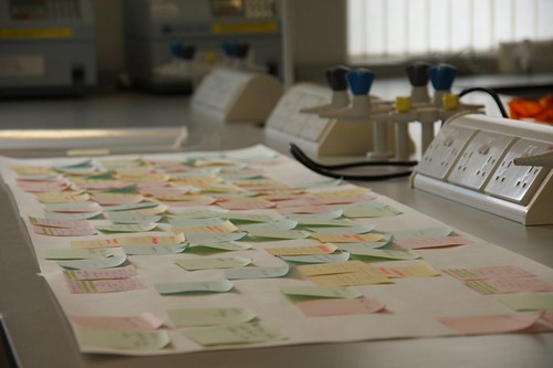
The post-its of failed ideas

One of the hardest parts of it, is trying to come up with what my PI called 'the pitch', the idea that is going to explain your project, why you are doing it, what it's for and (unfortunately seems to be a requirement) how it's going to save the world. The problem is that there are ~150 other teams also trying to think of a pitch, and there's quite a bit of pressure to find something new and original.

This might change as the competition gets bigger, it might become more acceptible to work on smaller ideas like "making a better assay for such and such" or "improving on the work done by a team last year". This I think would be an improvement, smaller ideas have a greater chance of actually working. Many of the smaller and less well funded teams have embraced this, and have very simple and succinct project ideas (i.e "biobrick protein A, put it into E. coli and test it").

Despite not being involved it is still fun to watch the work from the sidelines, and it means I have more time to look through what other teams are doing and to look at the process of iGEM more objectively. I might put up a few more posts about it over the summer (any time I'm too busy to go through papers probably!) but if you want to watch their progress I'd suggest following them, and watching the whole drama of lab work through undergrad-project eyes.

This is there project as well. They have help when they ask for it, and supervision when required. But mostly they've been just thrown into a lab with a load of equipment just to see what happens...